Newsroom
Advanced Imaging, Improved Ergonomics.
BRAINLAB MICROSCOPE INTEGRATION
Recognizing that neurosurgeons perform most of their procedures while looking through a microscope, Brainlab integrates with all major neurosurgery microscope vendors for advanced imaging and improved ergonomics allowing surgeons to stay focused on the surgical task.
With a dedicated marker geometry attached, the microscope is tracked by the navigation system like any other instrument, providing valuable benefits:
- Positional Tracking: The microscope’s focal point serves as the tool tip displayed on the patient data set, potentially replacing the use of the pointer
- Heads-Up Display(HUD): Displays all relevant information and objects – such as pre-planned tumors or fiber tracts – in the microscope oculars
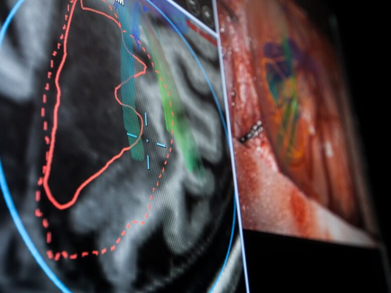
- Video Overlay: Reversely, all information seen through the oculars can be shown on the navigation screen as video overly
- Microscope Depthview: The reconstruction of the original MR or CT data according to the focal plane allows to continuously compare the live image with pre-operative data
- Remote Navigation Control: Using the microscope handles for improved ergonomics to control the HUD, take a screenshot or close the shutter