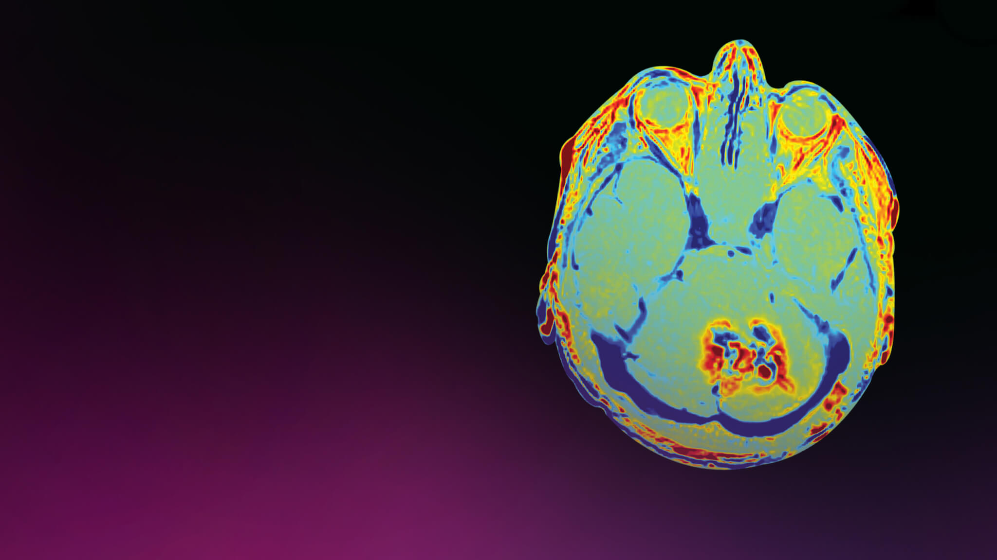
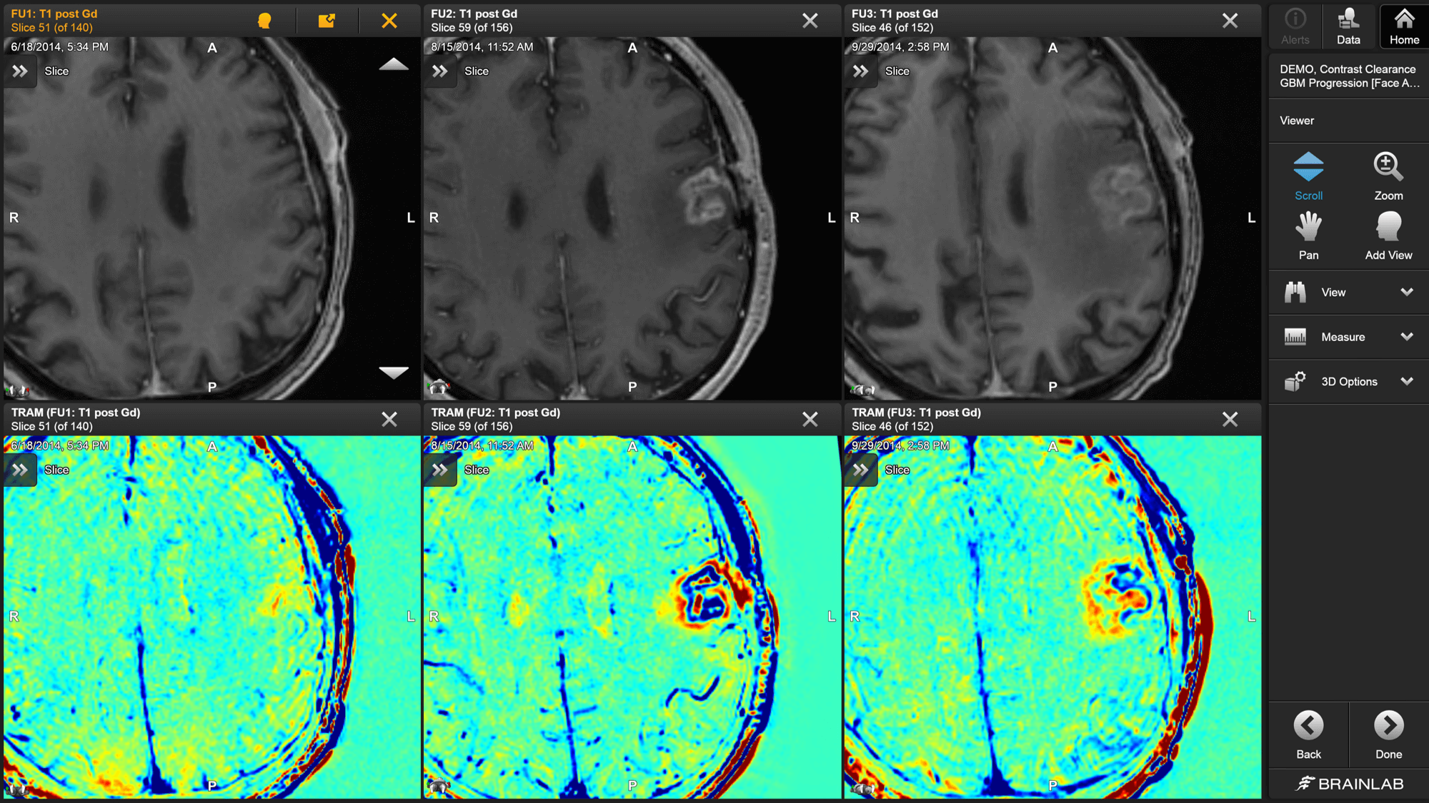
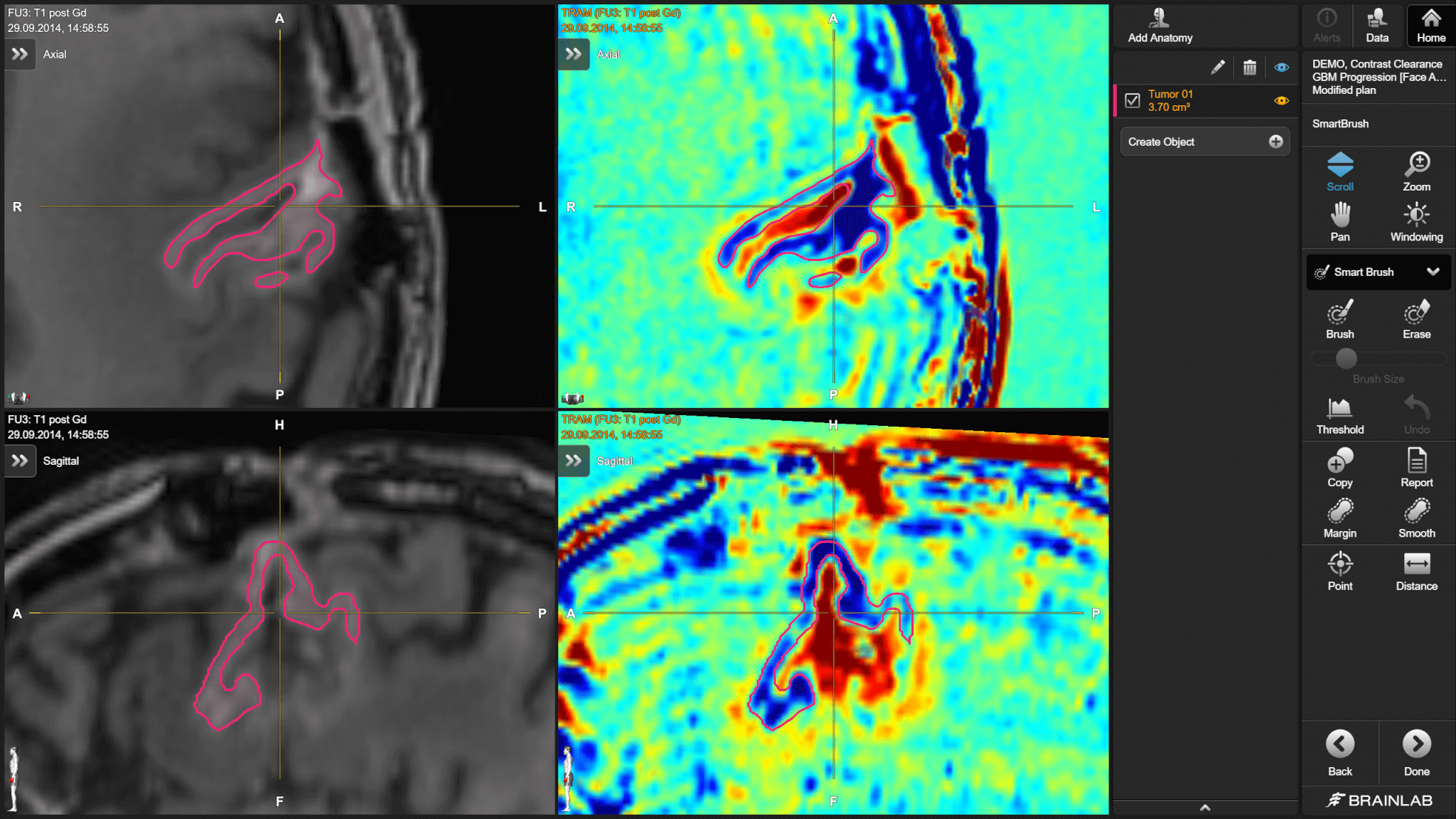
Elements Contrast Clearance Analysis is an MRI-based methodology for the differentiation of contrast clearance and accumulation regions in brain tumor datasets. The high-resolution analysis results provide additional insights to support ongoing assessment and decision-making.
Gain insights into tumor characteristics
Differentiate contrast clearance and accumulation areas with high sensitivity and specificity, enabling precise1 identification of tumor recurrence versus radio necrosis after radiosurgery.
Make optimal treatment decisions
Access additional information to support ongoing assessment of tissue characteristics during follow-ups and confident decision-making throughout the patient journey.
Support disease management workflows
Track patient progression using longitudinal data to detect therapy failure earlier and determine when new interventions are needed.
Turn insights into action
Discover how advanced post-treatment assessment tools can help you make confident decisions for optimal patient care
Developed for diverse clinical specialties
Elements Contrast Clearance Analysis, developed at Sheba Medical Center in Tel Aviv, serves multiple disciplines, including radiosurgery, radiation oncology, neurosurgery, neuro-oncology and neuroradiology.
Revolutionize your treatment decisions
Experience how Elements Contrast Clearance Analysis can support confident, data-driven treatment decisions that elevate patient care
Ready to add valuable information to your post-treatment assessment?
Bodensohn, R.; Forbrig, R.; Quach, S.; Reis, J.; Boulesteix, A-L; Mansmann, U. et al. (2022): MRI-based contrast clearance analysis shows high differentiation accuracy between radiation-induced reactions and progressive disease after cranial radiotherapy. In ESMO open 7 (2), p. 100424. DOI: 10.1016/j.esmoop.2022.100424
Source: Novalis Circle 2023 - The use of Contrast Clearance Analysis Software to differentiate Brain Tumors from Radionecrosis: A Revolution?


