keyvisual_cranialrt.jpg

Elements Solutions for Cranial SRSSimplify complex planning for cranial indications

Discover more

Elements Solutions for Cranial SRS simplify complex planning routines for cranial indications, such as pituitary adenomas, vestibular schwannomas and arteriovenous malformations (AVMs). A dedicated 4Pi arc setup optimization and a VMAT algorithm designed for cranial radiosurgery provide improved plan quality and MU efficiency at the same time. 

Add consistency to all your plans

Minimize intra- and inter-observer variability compared to manual outlining with our automation-supported tools. Ensure consistent, time-efficient image co-registration and segmentation of targets and organs at risk with RT Elements, forming a reliable foundation for creating high quality dose plans that fulfill defined treatment criteria.

Create high quality SRS VMAT plans

Elements Cranial SRS combines template-based treatment planning with a dedicated 4Pi arc setup optimization algorithm to ensure high quality treatment plans with minimized modulation for robust dosimetric results and MU efficiency.

Achieve submillimetric accuracy

Maintain low margins and treat with fewer fractions using Elements Cranial SRS in combination with ExacTrac Dynamic during treatment delivery. Improve patient experience and drive program throughput with Brainlab RT portfolio. 

Get to know our indication-specific Elements

Create high-quality SRS plans easily and effectively for single cranial targets with protocol-based VMAT radiosurgery planning. 

Elements Cranial SRS in 3 minutes

Preparing for cranial planning

We’ve built automation into every step of the pre-planning workflow, including image fusion, distortion correction, object outlining and segmentation. Save time with fast and easy-to-use tools, add consistency and gain confidence from the submillimetric accuracy you’ll achieve along the way.

Rigid and deformable co-registration

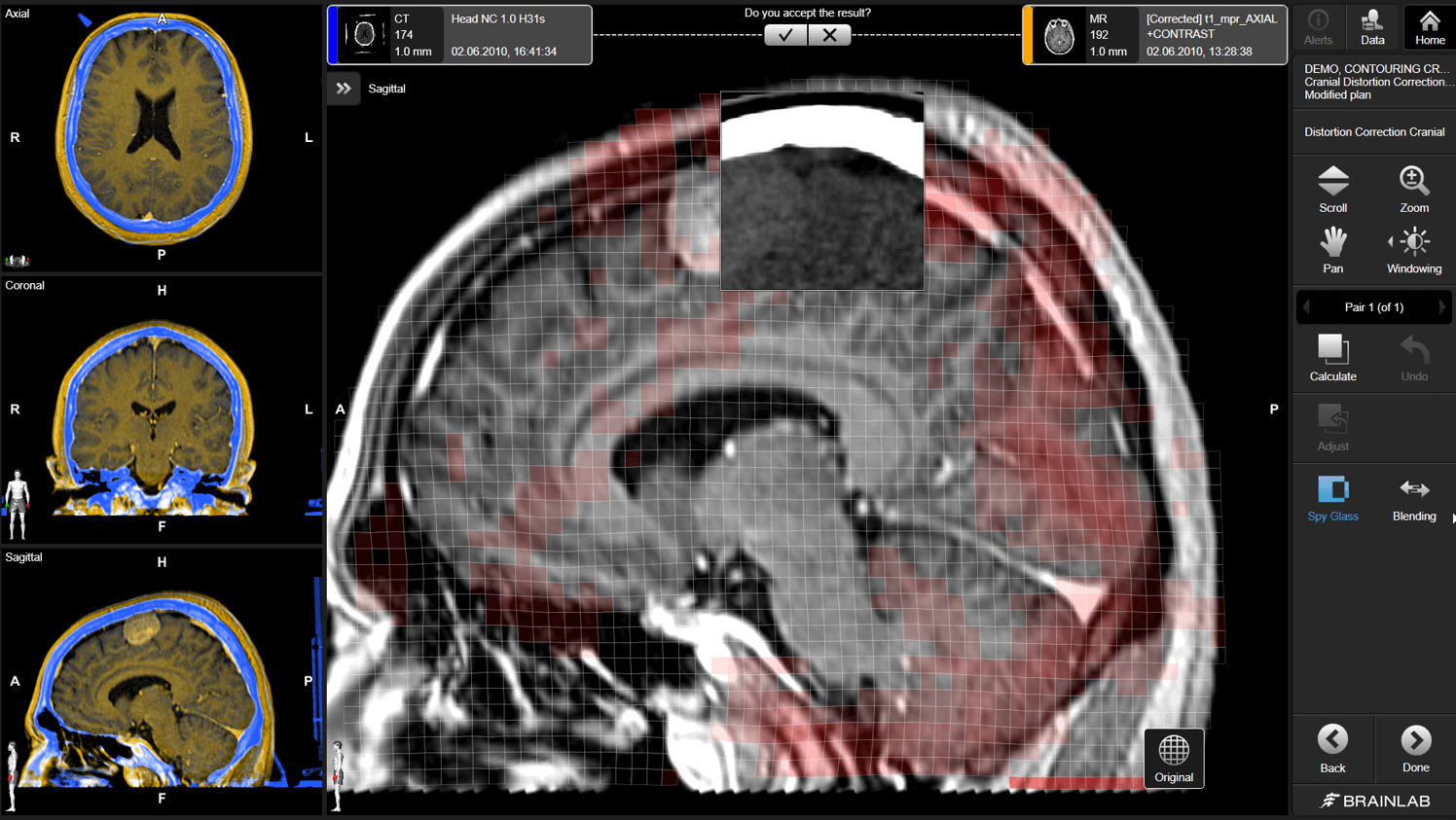
Elements Distortion Correction Cranial corrects patient-specific imaging distortions by applying a deformable registration that respects the anatomical ground truth. 

Screenshot of Elements Distortion Correction: correction of fused images

Organ-at-risk segmentation

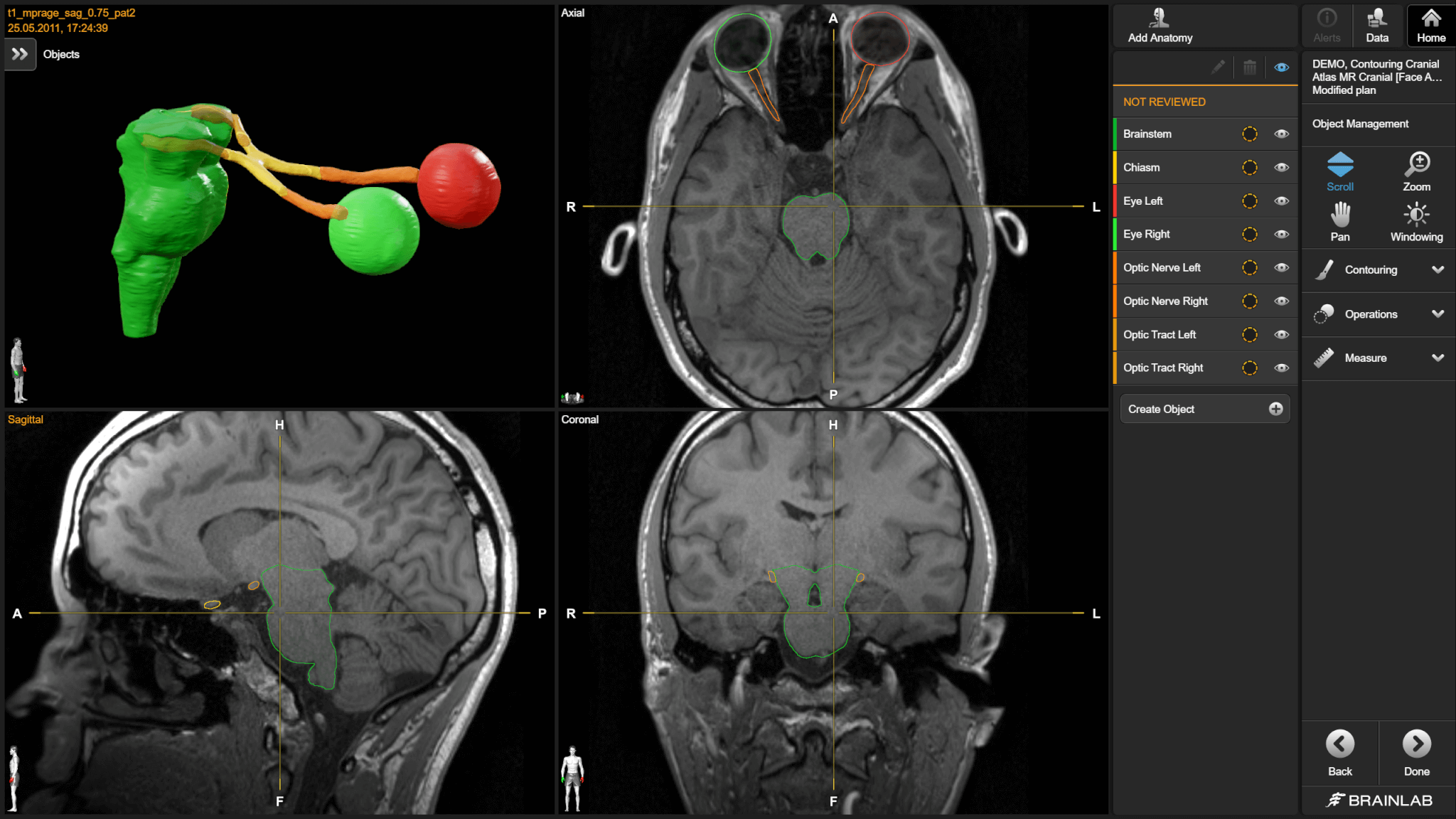
Elements Anatomical Mapping automates the time-consuming process of contouring organs at risk by efficiently, consistently and accurately registering datasets to a synthetic MRI-based tissue model. The tissue model includes more than 100 cranial structures.

Screenshot of Elements Anatomical Mapping: organ-at-risk segmentation

3D target outlining

Elements SmartBrush defines volumes using only two slices. This application outlines cranial tumors quickly and easily on multiple modalities, minimizing intra- and inter-observer variability compared to manual outlining. Once delineated on two slices, the full volume is automatically generated and associated reports can be produced.

250407_aiseg_banner_lum_1.png

Elements AI Tumor SegmentationEnhance cranial tumor delineation
with AI-powered tools

Elements AI Tumor Segmentation can segment supported cranial tumors
in multiple brain mets, cranial and retreatment review workflows.

Experience cranial SRS planning with Elements Radiosurgery Solutions

Creating the dose plan with Elements Cranial SRS

With pre-planning complete, dose planning with Elements Cranial SRS is the next step. Easily and effectively create consistent, high-quality SRS plans for single cranial targets with protocol-based VMAT radiosurgery planning. 

Patented 4Pi arc setup optimization

The 4Pi optimization algorithm, combined with highly conformal VMAT-based dose delivery designed for SRS, makes it possible to avoid critical structures while enabling high quality, efficient plans.  

Unique 3D views

Dose interface and unique 3D views highlight the most relevant dose coverage metrics and allow a comprehensive assessment of dose distribution. 

Robust dosimetric results

Minimized modulation balances MU efficiency with target coverage for robust dosimetric results and efficient delivery times. The adaptive dose grid automatically provides additional calculation points for small targets based on structure size. 

Highly precise beam modeling and dose calculation

By using Brainlab reference beam models, the measurements for the highly accurate Monte Carlo dose calculation algorithm are reduced by 90% for all TrueBeam® platforms with HD120™ or Millenium MLC with 6MV and 10MV, standard and FFF mode, as well as Elekta Versa HD™ (Agility™ MLC) with 6 MV and 10MV, standard and FFF mode.

Click on the link below to learn more about how the Brainlab reference beam models were developed and validated. 

Icon Set: Measure, Select, Plan

Related Products

Related Information

Innovation isn’t often free, but our demos are

Find your future workflow today.

  1. Source: ASTRO 2019 Novalis Circle Symposium: Initial Clinical Experience: Elements Multiple Brain Mets SRS  

  2. Source: Novalis Circle 2023: Elements Cranial SRS – 4Pi Optimisation case studies.