
Integrating confidence in real time.
We transform intraoperative ultrasound into a powerful navigation tool to support maximum safe resection, even when brain shift, registration or navigational inaccuracies occur.
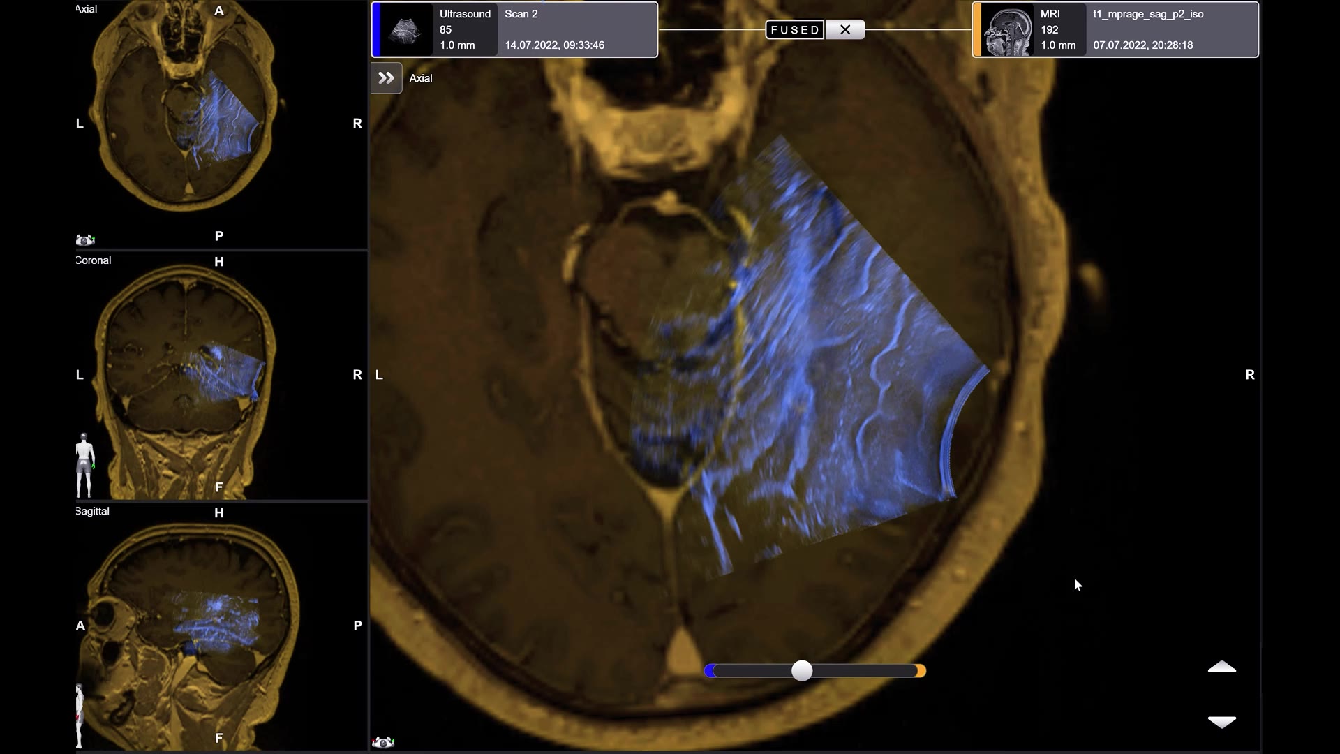
Real-time correlation of intraoperative ultrasound and preop MRI
Even with advanced navigation and registration methods, loss of navigational accuracy can occur during neurosurgery. With Ultrasound Snap to MRI, you can quickly and reliably realign your preoperative MRI, helping to restore navigational accuracy and providing a strong foundation for confident surgical calls.
Precision and confidence in navigating cranial tumor resections
Enhance your surgical precision with advanced navigation accuracy that supports even complex tumor resection procedures, such as posterior fossa cases. Detect and correct registration errors or loss of navigational accuracy in real-time, ensuring maximal safe resection and reliable identification of residual tumor.
Efficient intraoperative imaging without workflow interruption
Leverage fast, cost-effective intraoperative imaging that integrates seamlessly into your surgical routine—providing real-time insights and helping maintain navigational accuracy throughout the procedure, without disrupting your workflow.

Research that’s making waves in neurosurgery
Join the wave
Get the navigational accuracy you need to achieve your goals.

Peel back the layers of Ultrasound Navigation

Take a deep dive
Check out the real-world evidence you need to make the shift.


Your new intraop imaging journey starts now
Maximize insights with science-backed Ultrasound Navigation.