microscope_navigation_spine_keyvisual_16to9.jpg

Microscope Navigation for Spinal WorkflowsClarity in every layer

Discover more

Surgical clarity, redefined for spine. Microscope Navigation enriches your field of view with augmented reality by projecting patient-specific anatomical objects into the microscope oculars. These real-time overlays are integrated with navigation to support intraoperative orientation and guidance in narrow surgical corridors, while preserving surgical flow.

250915_microscopenavigation_spine_benefit1_orientseamlessly.jpg

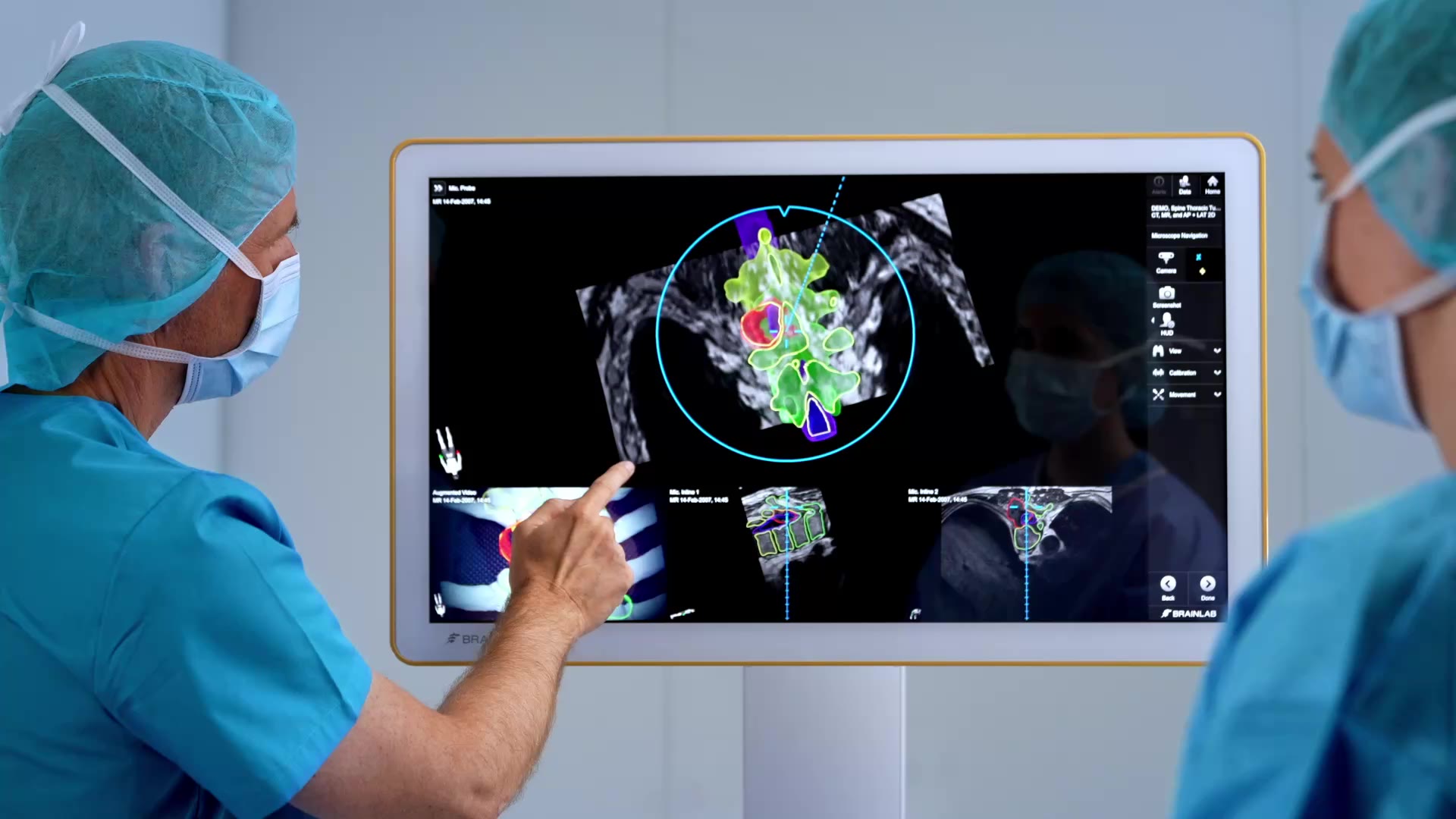
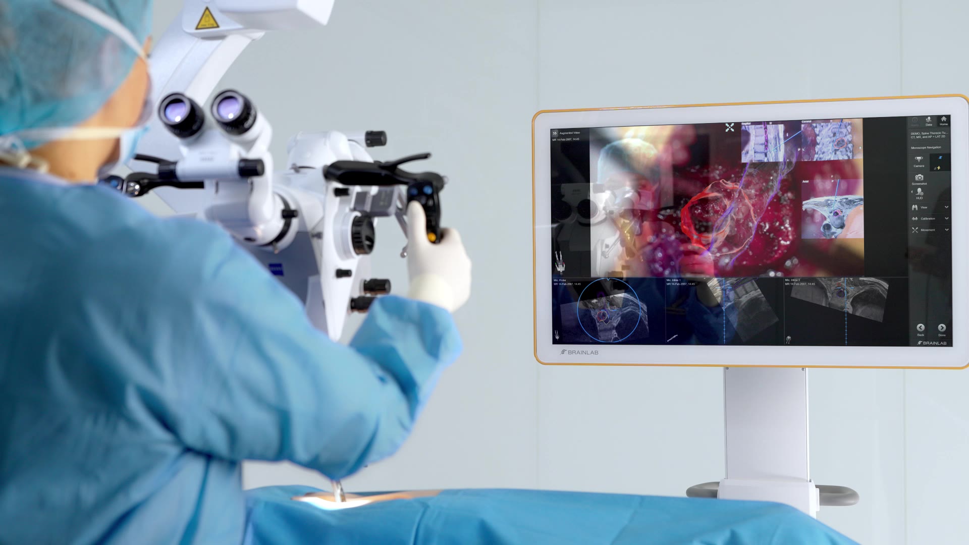
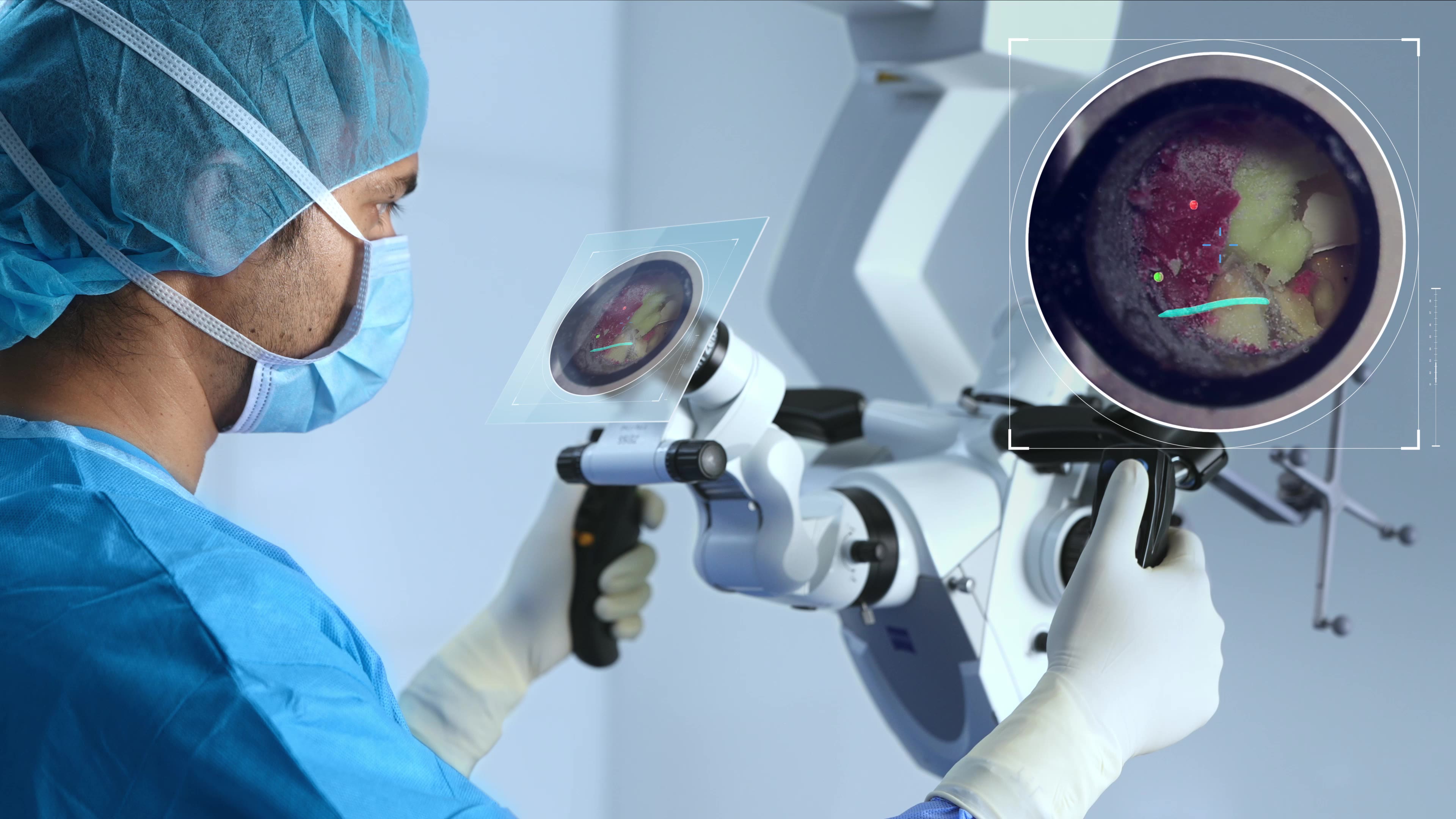
Orient seamlessly approach confidently

Stay oriented, no matter the approach. Augmented reality overlays, fully integrated with real-time navigation, support fast anatomical recognition and smooth adaptation to microsurgical workflows—empowering both experienced and emerging surgeons to navigate complex anatomy with assurance.1

250915_microscopenavigation_spine_benefit2_visualizebeyond.jpg

Visualize beyond the visible

Reveal critical anatomical structures like nerves, vessels, tumors and hidden bone landmarks—often obscured in confined surgical spaces. Augmented reality overlays derived from Elements Object Management enhance intraoperative visualization, providing deeper anatomical insight and helping you operate with confidence during complex spinal procedures.1

250905_microscopenavigation_spine_keybenefit3.jpg

Stay in flow stay in control

Focus fully on the patient and procedure. Step-specific anatomical information appears directly in the microscope oculars, so you stay aligned with the surgical plan without shifting attention to external screens. Whether performing a TLIF, spinal tumor resection or decompression for stenosis, maintain smooth workflows and precise control from start to finish.2

Augmented reality in spine surgery will significantly improve the way that we take care of patients, not only for instrumented spine surgery, but also for decompression surgery and pain procedures.

Roger Hartl, MD, Neurosurgery Spine, Weill Cornell Medical Center, New York, NY, United States
micr_spine_kol_hartl_16x9.jpg

The ability to toggle through grouped anatomical objects in real time has transformed how I approach spinal tumor cases. It’s like having a dynamic roadmap that adapts to each step of the procedure streamlining my workflow while keeping me focused, informed, and in control throughout.

Marcel Ivanov, MD, PhD, Department of Neurosurgery, Royal Hallamshire Hospital, Sheffield, United Kingdom
micr_spine_kol_ivanov_16x9.jpg

Integrating augmented reality with microscope navigation revolutionizes surgical procedures, enabling surgeons to achieve improved visualization and orientation, thereby significantly enhancing our surgical capabilities in the care of our patients.

Adetokunbo A. Oyelese, MD, PhD, Spinal Surgery Division, Brown University Health, Providence, RI, United States
micr_spine_kol_oyelese_16x9.jpg

Integrating augmented reality with microscope navigation is transforming how we teach and learn in the O.R. It makes complex anatomy more intuitive, accelerates the adoption of minimally invasive workflows and ultimately helps build confidence faster.

Andreas Kramer, MD, Neurological Surgery Department, LMU Klinikum, Munich, Germany
micr_spine_kol_krammer_16x9.jpg

Bring anatomical insights into view—without breaking surgical flow

Discover how Microscope Navigation supports orientation and decision-making in even the most complex spine cases.

Intelligence in your field of view

Augmented reality is becoming essential in spinal microsurgery, where limited visibility and complex anatomy demand precise, real-time anatomical orientation. With Elements Object Management, you can preplan and segment anatomical structures, like tumors, target levels for TLIF or areas of stenosis, and then visualize them as semi-transparent 3D objects over the native anatomical view with Microscope Navigation. During planning, anatomy can also be explored from multiple angles and planes, revealing hidden structures and offering deeper insight into complex regions.

spine-microscope-navigation-feature-1-int-fieldofview-mp4

Uninterrupted focus

Microscope Navigation is designed to keep you focused on what matters most—your patient. By integrating real-time anatomical and instrument visualization directly into the microscope view, there is no need to look away from the operative field. Ergonomic visualization tools support continuous focus:

  • Adjustable augmented overlays provide clarity without visual overload

  • Integrated navigation views support spatial orientation even in low-visibility conditions

  • Hand grip controls allow you to access navigation and augmented reality overlays without breaking concentration1

spine-microscope-navigation-uninterrupted-viewing-mp4

Step-by-step guidance for microsurgical workflows

Microscope Navigation mirrors your surgical procedure by aligning visualization with each step of your workflow. Predefined anatomical landmarks or grouped objects appear as targeted augmented reality overlays, guiding you step-by-step through a concrete microsurgical workflow in spinal surgery. With intuitive handle control, toggling through the grouped objects and navigation views is seamless, allowing you to focus on the individual steps of the procedure. This step-by-step guidance also makes it a powerful learning tool for emerging surgeons, offering clarity and consistency in complex spinal microsurgeries.2

spine-microscope-navigation-feature-3-misworkflow-mp4

Hands-free alignment with robotic control

Microscope Navigation enables robotic positioning that’s ergonomic, efficient and intuitive. Instead of manual adjustments, you can rely on:

  • Continuous autofocus to a predetermined point of surgical interest

  • Automatic return to a predetermined anatomical landmark

  • Automatic alignment of the microscope to a navigated instrument or instrument tip

250915_microscopenavigation_spine_feature4_roboticcontrol.jpg

Freedom of choice

The use of microscopes in the O.R. is evolving, and Brainlab solutions adapt with it. Whether you prefer the ocular view or the exoscopic approach with video images displayed on large 3D monitors, Microscope Navigation supports both workflows. Exoscopic spinal navigation offers:

  • Ergonomic design that helps promote good posture and reduce fatigue—especially during long, demanding spinal surgeries—helping you maintain focus throughout the procedure

  • Shared visualization that immerses the entire surgical team in real-time view of the operative field, improving communication, coordination and teaching opportunities3

250915_microscopenavigation_spine_feature5_freedomofchoice.jpg

Curious how we can support your microsurgical cases?

See how Microscope Navigation integrates into your surgical workflows. From preplanning to intraoperative alignment, our specialists can walk you through your options.

Use cases for Microscope Navigation

Microscope Navigation helps you stay focused on what matters—bringing patient data directly into view.

Precise visualization for spine surgery

Microscope Navigation for spinal workflows supports a wide range of microsurgical procedures across the cervical, thoracic and lumbar regions. From degenerative cases such as TLIF and decompression for stenosis to complex spinal tumor resections, it provides precise guidance throughout your surgical workflow.

250915_microscopenavigation_spine_usecaseimage.jpg

Focused on precision for cranial tumor resection

Microscope Navigation supports tumor resection by delivering real-time, data-driven guidance directly into the surgeon's line of sight. With in-line augmented reality integrated into the microscope optics, Microscope Navigation helps identify tumor boundaries, distinguish critical structures, and adapt to brain shift—supporting safe resection while preserving healthy tissue.

250915_microscopenavigation_cranial_benefit3_realignconfidence.jpg

Related products

Proven impact in the O.R.

  1. Screenshot courtesy of Marcel Ivanov MD, PhD

  2. CE mark available. FDA 510(k) pending. Not yet commercially available in several countries. Please contact your sales representative.

  3. Screenshot courtesy of Miriam Bopp, MD, PhD