Are you in %country%? Do you want to visit our website in another language?

Webinar

Combined Approaches in Frontal Sinus and Skull Base Surgery

Topics
Surgery Navigation
ENT
Language

English

Share this page

Description

Brainlab invites you to join our live webinar, “Combined Approaches in Frontal Sinus and Skull Base Surgery”, on December 9, 2020 at 4:00 pm CET presented by Prof. Dr. med. Fabian Sommer, Consultant at ENT department, University Hospital Ulm, Germany.

This webinar will cover topics including:
• Frontal sinus and skull base pathologies with combined approaches
• Intraoperative navigation and its role due to proximity to cranial base and orbit
• Inverted papilloma of the frontal sinus, ngocele/meningoencephalocele, defect reconstructions of the frontal skull base by pericranial flap and “mailbox approach”

We look forward to meeting you online!

Language | English

In case you can not join the webinar, it will be recorded and shared afterward.

Participation is free of charge.

Speaker:

Prof. Dr. med. Fabian Sommer

Consultant at ENT department, University Hospital Ulm, Germany

Video Transcript

Jana: Welcome to our second webinar in our ENT webinar series. My name is Jana Neider and we are live from the Brainlab Tower in Munich in Germany. It’s a pleasure for us that you are here with us today.

Before I introduce you to our speaker, Professor Sommer, I would like to explain a few points. This webinar will focus on the combined approaches in frontal sinus and skull base surgery. The lecture will last about 45 minutes followed by a question and answer session. Questions can be only submitted through the online chat function. Those questions will be selected by myself and addressed to Doctor, Professor Sommer after the meeting. The webinar is live, but it will be recorded to be watched again at any convenient time for you. After this meeting you will receive a special link to have the recording available. For further questions after the webinar, please feel free to use our online chat function.

Now onto our speaker, Professor Sommer. I’m very happy that you are here today. Professor Sommer is an ENT consultant at the ENT department at the University Hospital of Ulm in Germany. He is a member of the European Rhinology Society and highly interested also in new technology. That makes him an ideal speaker into our webinar series as well as a very experienced surgeon in the global ENT community. We are very much looking forward to have you here today speaking about your experience. Professor Sommer, the virtual stage is now yours.

Prof. Sommer: Thank you very much. I’m just making my presentation ready.

Jana: Thank you, Professor Sommer. Perfect.

Prof. Sommer: So there we are. I hope you can see everything on the screen. Thank you very much, Ms. Neider, for the kind introduction, and it’s a pleasure to be part of the webinar series. It’s a pleasure to report about our experiences in combined approaches in frontal sinus and skull base surgery. The following presentation is based on the experiences that we made within the last few years, and I will highlight some cases that we operated on to enlarge the picture that I would like to depict for the indication of combined approaches.

The presentation is divided in three parts. I will start with frontal sinus surgery and the combined approaches in this area, and then head over to skull base surgery, and in the last part I would like to focus on intraoperative navigation and some aspects that might be helpful especially in skull base surgery.

Coming to frontal sinus surgery. In our department we do not address the frontal sinus in ever paranasal sinus surgery. The patient should have specific complaints or have a specific reason that makes us enter the frontal sinus or the frontal recess area. As for an example, feeling of pressure in the forehead region or headaches. This is very often the case in revision surgery, and in revision surgery sometimes a combined approach is necessary. Basically there is no “regular” frontal sinus surgery because the anatomy is highly variable, and you all know that there are a number of variations that can happen. I would like to focus on that a few slides later.

For addressing the frontal sinus, some preparations are important. First of all, the informed consent. It is mandatory to discuss with the patient every possible approach besides the front nasal approach. Depending on the pathology that is addressed, an open approach might be needed, and this must be discussed with the patient prior to the operative procedure. Second, a CT scan is mandatory, or an MRI scan in some cases is okay as well, but in general it is important to decide prior to the procedure, do I need navigation? And is the data set that I have available for navigation? Last, but not least, a CT checklist should be filled in, and I would like to come today’s anatomic variations that are included in the CT checklist in two slides.

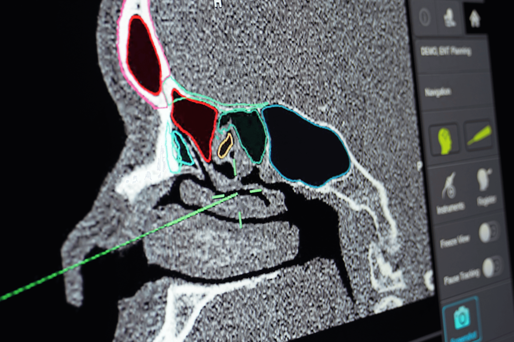
We use a Brainlab navigation system. We have the Brainlab Curve system. It’s an optical system. We are very happy with that. There are different systems on the market, like optical or electromagnetic systems, and also combinations of both are available. Each system has advantages and disadvantages, and in general one has to find the optimal system for the kind of surgery that is planned.

Coming to the CT checklist. This checklist should include the anatomic variations of the uncinate process because these variations determine the way up to the frontal sinus and the frontal recess. So if we have an insertion, like here on the left side of the patient at the skull base, we have the entrance to the frontal recess on the left side of it, so on the lateral side. And on the right side of the patient we have a so-called terminal recess, and the frontal recess will then start medial to the uncinate process.

It should contain the variations of the cranial insertion of the middle turbinate as this insertion is located directly at the skull base, and the cribriform plate in this area is very thin and very, very fragile, and this must be taken into account. The ethmoidal bulla has some anatomic variations as a supra bulla cell or a supra bulla frontal cell that might make the way into the frontal sinus a little bit more difficult.

Configuration of the lamina papyracea or protruding into the ethmoid sinus protruding orbit, which is sometimes a little bit bigger. The configurations of the olfactory fossa, especially the Keros type three, when the olfactory bulb is coming way, way down into the frontal ethmoid region.

And last, but not least, the AP diameter between the skull base and the frontal beak because this determines the width of the entrance into the frontal sinus or the width of the frontal recess. And this also determines the kind of approach in some cases and the instruments that I have to select. Do I have the correct instruments to enter the whole frontal sinus or the pathology that is located in the frontal sinus in a transnasal route on its own? Or do I need a combined approach?

In 2016 the International Frontal Sinus Anatomy Classification was published, and this classification made a nomenclature of the variations a little bit easier. It is determined between the agger region and the bulla region and the in-between lying frontal recess. And several anatomic variations in the recess area can occur like a supra agger cell, a supra agger frontal cell with an extension into the frontal sinus, or a bulla cell, like a supra bulla cell, and a supra bulla frontal cell that extends into the frontal sinus with a direct contact to the skull base which is sometimes important if you are planning to enter the frontal sinus in a transnasal route.

There are also different possibilities to perform an open approach. First of all, the eyebrow incision, which we use in smaller pathologies, but on the other hand there is so-called bicoronal incision which gives you the most extended approach and the best overview because all of the anterior of the frontal sinus can be removed, and the visibility is very good in that case. But there are also modifications like the transpalpebral orbitofrontal approach which was published in 2017 in “Laryngoscope.”

Now I would like to present you some examples that we operated on to highlight what we think possible indication for a combined approach might be. The first case is something that you all might already have seen. It’s a frontal sinus opacification on the right side. It’s visible here. The patient presented with swelling of the eye and with feeling of pressure and sometimes headache in that area. And it became clear that he already had paranasal sinus surgery. Obviously it came to the formation of a mucocele in the right frontal sinus that already destroyed the base or the ground of the frontal sinus at the border to the orbit.

Prior to the procedure it is important to think about the correct way to address that pathology. Is it possible to remove the medial wall and to remove more than just a small hole? To make a big opening in a transnasal route? In this case, it is definitely possible. This is a typical mucocele, which is easy to operate on, quite easy. You see the frontal recess. This a 45 degree optic endoscope, and you have to look up into the frontal recess. The mucocele is inside, and you see the secretion coming out. And then the second very important step is to widen the opening as far as possible so you have a direct view into the frontal sinus afterwards. So this is a case that is definitely operateable in a direct route to the transnasal approach.

In the patient that we have here, it’s a patient also with a mucocele formation on the right side. This patient also has adhesions in the bony border to the orbit, and the right frontal sinus is opacificied. But when having a look at the more posterior CT scans and coronal view we see that the frontal recess is quite narrow, and it might be very difficult to enter that area to remove the medial wall to create a big opening in that patient.

And in these patients it is sometime helpful if adhesions already exist, like in this area, in the anterior superior orbital rim, because in this case we can just make an incision and enter the mucocele from an open approach and have a look into the mucocele, which is visible now. You see that the secretions always suck out. We see here the entry to the frontal recess, which is completely scarred. But the mucosa itself looks quite nice. That’s okay. Coming back to the frontal recess area there is no rest opening, so it’s completely locked.

Now we can combine both approaches. We enter from the nose. This is a 70 degree endoscope, and we have here the axilla of the middle turbinate. The frontal recess is already widened. Now we can have a look into the narrow frontal recess that I just showed you in the posterior CT scans and can identify the medial wall of the mucocele by palpation from outside and protect the skull base which is directly behind. And after resection of the whole medial wall, we have a wide opening in the the frontal recess and can see the suction coming from the transnasal approach. So in this case we have an opening that should stay open for a long time.

Coming to the third patient, which is even a little bit more difficult. It’s a patient with a history of a car accident and multiple body fractures in the mid-facial area and the skull base. And as you can see, the frontal sinus on the left side is not just one cavity. There are different chambers, and that makes an approach quite difficult. When having a look at the CT scans, it might make you think that this is operateable or addressable in a transnasal route, but the lateral aspect and the lateral chambers are sometimes very difficult to address or to reach with the instruments in a direct route in a transnasal approach.

And also, the frontal beak is very prominent in that patient, so this additionally makes the entrance into the frontal sinus a little bit more difficult. Coming to the procedure, this is the endonasal approach. We see the middle turbinate, the resection of the uncinate process, and the frontal recess is prepared. First of all, the lumen is identified, and then we prepared, and then we switched to the external approach.

We see here the eyebrow incision, preparing through the periorbital muscle, and we had a small opening which was enough for entering the frontal sinus with an endoscope, but widened it a little bit to be able to enter with an endoscope, and additionally with an instrument. And now this is the endoscopic view into the fully scarred frontal recess. It can be identified by means of navigational aid. After identifying it, the membranes are removed. And now it is important to use the drill to get an opening to the frontal recess and the nasal cavity, which should be as big as possible.

And in my opinion, in some cases, the combined approaches offer the possibility to use, for example, a burr from below and to control what you are doing from above. I think you all already had the situation when working with a 70 degree endoscope and additionally having the burr inside is sometimes very difficult to handle, and in this case it is a little bit easier because you can control from the outside what you are doing from the transnasal route. After widening the frontal recess, it is important to close the defect. You can either use PDS foil, or in most of the cases we use ear cartilage if there is an opening which is more than, let’s say, four to six millimeters, and the rest doesn’t have to be reconstructed.

Coming to the last patient of frontal sinus surgery, that was a case where we operated in an inverted papilloma of the left frontal sinus. And in these cases it is very dependent on where the inverted papilloma is located, where the origin is. In that patient was located very far lateral, so the transnasal approach was not enough, so we had to switch over to a combined approach. And again, we did an eyebrow incision and entered the frontal sinus through a kind of mini-trepanation with an endoscope and an instrument to remove the mucosa in that area. It was a combined approach, and the surgery went very well.

And I would like to show you a short video of the regular follow-up. These patients usually come every six to eight weeks after the surgery in our hospital to have a follow-up, and this follow-up is one year after surgery. You can see the nasal cavity and the view up to the frontal sinus. This is a flexible endoscope because it allows you to enter the front sinus in total. And you can see the screws in the anterior wall of the frontal sinus, which fixate the lid that we created, and even the lateral and cranial aspects of the frontal sinus look very well.

So in these patients we have an opportunity, if the frontal recess is open, to enter the frontal sinus and to have a look and to have a close follow-up just to see if there is a recurrence of the inverted papilloma. And this is very important because in up to four percent of the people with inverted papilloma there is the possibility of a malignization of this entity. So it must be taken into account that the patient has a possible mutation into a malignancy, and that’s why we are very keen on having close follow-ups.

Summing up combined approaches and a short look at the current literature. Some manuscripts and publications focus on frontal sinus CSF leaks and postulate that the combined approaches have better results in that area, especially in the frontal sinus, because the exposure is better, the success rate is supposed to be way better than just using a transnasal route.

Coming to the inverted papilloma there is one interesting study from 2017. They had four cases with an inverted papilloma of the frontal sinus that was resected in a combined approach. And they had, in all these cases, during a period of four years, no recurrence, which is, I think, good. It’s not really many cases, I have to admit, but it is hard to find. There are no studies that have way more cases with that in the frontal sinus.

In our experience, combined approaches are very useful when having extensive pathologies, especially in post-traumatic pathologies, if there are fragments and big formations of scar tissue or if the exposition in general, the pathology is very difficult. Two weeks ago we had a patient who had a lymphoma, or was suspected in the MRI scan to have a lymphoma on the very lateral aspect of the frontal sinus. And in that patient, it was also not possible to enter the frontal sinus is a transnasal route. Or it was possible to enter, but it was not possible to get a biopsy from the lateral aspect, so we had to switch over to a combined approach. And we did also an eyebrow incision.

So in this case we had a difficult exposition, especially for getting the biopsy. And generally speaking one should have a plan prior to the procedure, but there is no master plan before every procedure. And one has to be aware of the fact that you might change the way that you approach the pathology.

With that, I would like to head over to skull base surgery. It was in 1993 when a report from the German Federation of Oto-Rhino-Laryngology head and neck surgery was published. In this report we had description of typical approaches for frontal skull base malignancies. And there we have the lateral rhinotomy, we have the subfrontal approach, the transfacial approach, and the midfacial degloving. That’s what I’m looking for, midfacial degloving approach. These approaches were discussed as very modern ones and very effective ones.

Within the last 20 to 30 years there has been a change in technique, technical equipment like navigational systems and better instruments, better optical systems. We now have 4K endoscopy, which enables us to resect more clearly and to operate through narrower holes. Nowadays, when we speak about skull base surgery, the main indications are tumor, trauma, and malformation, and there are important prerequisites or requirements prior to every surgical procedure.

The first of all is that there is a team of a neurosurgeon and ENT surgeon that are experienced in working together. It should not be the first case that they operate together. They must be used to each other and must know what to do in specific situations at the skull base. And it is also very important to have neuroradiology in the house just in case there is a major bleeding, for example the internal carotid artery, for the case that the bleeding is not stoppable in an operative way at once.

Hardware requirements should be endoscopes in any thinkable angulation. We usually have 0, 30, 45, and 70 degrees available. We have two 4K endoscopies in our OR, but it should be at least full HD or HD resolution. Long instruments are necessary to enter the intracranial space, especially behind the sphenoid sinus. And navigational system is mandatory. In some cases, intraoperative imaging can be helpful, especially in cases where we operate with the neurosurgeons together and where the main part of the pathology is located intracranially.

In our OR, in the ENT OR, we have a mobile, digital volume tomograph scanner. It’s a Xoran xCAT. We are able to perform a CT-like scan in a patient in general anesthesia. There are different options like here in the hybrid OR where we have also a digital volume tomograph scanner mounted on a robot industrial arm. And that allows us to perform scans in every thinkable position of the patient.

And last, but not least, we have the brain suite in the neurosurgical department. And there we can achieve an MRI scan in a patient during a procedure, and then integrate these pictures into the navigational system. It is sometimes recommendable to use intraoperative imaging, especially in bigger surgical procedures where the anatomy is changed in a dramatic way, to get new landmarks and to have a more detailed land map to operate on.

Coming to the first case, it’s a patient with a glioma that was operated in our department, and it was a patient 39 years of age. He had frontal completes, especially headache, frontally located. And we see here in the CT scan an opacification of the frontal sinus and recess area, additionally to a thinned looking bony border or bony skull base. And the MRI scan revealed intracranial mass protruding into the frontal recess and the frontal sinus. That’s what we were addressing in doing the procedure.

Coming to the surgical approach, in this case we chose a combined approach because sometimes it’s difficult to close the defect of the posterior wall in a transnasal route on its own. Here you see the resection of the glioma in the frontal sinus and the frontal recess region. And as you can see here we used natrium fluorescein to be sure that there is no CSF leakage after the procedure. And after removing all of the glioma we repaired or reconstructed the dura with a neuro-patch, TachoSil, then put on a galeal periosteal flap. And after that, the bony anterior wall of the frontal sinus can be replaced with titanium screws, and the frontal sinus is reconstructed.

Coming to case number two. It was a female patient who presented in our departments as well with headaches in the anterior left lateral portion. And in the CT scan we detected a big defect in the left frontal skull base, anterior skull base, with kind of tissue, intracranial tissue protruding into the ethmoid sinus. And also the MRI scan revealed that and made that more clear that we had a meningoencephalocele in that patient.

This pathology was…we discussed the correct approach. We decided not to use a transnasal route on its own because the width of the defect, as to be expected, would be too big. And in the lateral aspects we need some stable structures to put on reconstructive tissue. So we chose a combined approach to get in with a neurosurgeon.

This is the picture from the craniotomy where the frontal bone was opened and then finally resected. You see the frontal lobes behind the dura, and the frontal sinus is opened. And after a cranialization of the frontal sinus, the protruding mass was resected and prepared. You see the skull base, and these scar tissues and these small, yeah, bubble-like structures that are protruded brain mass. And after resecting the mass that entered the ethmoid or the nasal cavity, we are right at the posterior edge of the [inaudible 00:27:01]. And after resection we have a big defect going into the nasal cavity.

Now this is a view onto the nasal septum from above, and middle turbinate is located here. And now, in the next step, the reconstruction must take place. We decided to use a calvarial split graft because there was no bony structure, and the cavernous link grafts in the tabula externa gives a very good stability to that area, so there is no mass protruding anymore.

And then in the second step of reconstruction with a galeal periosteal flap is put inside which reconstructs the dura, the inner lining. Afterwards the bony anterior part of the skull is replaced with titanium screws. And in the last part of the procedure we reconstructed the outer lining or the border to the nasal cavity with a nasoseptal flap, which is prepared here with the electric knife, electric needle. The mucosa is prepared from the septal cartilage. And after putting it in the posterior part of the nose, it is rotated for 90 degrees and placed up to the skull base, fixated with fiber glue and TachoSil. And then a nasal dressing for five to seven days is put inside. And the procedure is two and a half years ago now, and the patient is doing very fine. She had no recurrence.

So the the technique for reconstructing bigger effects like in this case is a pericranial flap from above. Use a medial structure with a calvarial split graft. And then the final step is a nasoseptal flap from below, which is very stable and you could call in the kind of work house for the bigger defects and nasal reconstruction because it’s very, very stable and has a very low rate of post-operative CSF leaks.

The next case is patient that was operated in an external hospital and presented in our department with acute epistaxis. This patient had a history of adenoidcystic carcinoma in the posterior ethmoid and sphenoid sinus. He received a transnasal resection one and a half years ago and underwent radiotherapy with a heavy-ion boost. And when coming to our department, the bleeding immediately stopped which was very interesting. We then performed a CT angiography, and, you might have suspected it, in the right part of the sphenoid sinus we have adhesions of the bony and mucosa wall of the internal carotid artery, which finally led to bleeding. Luckily the bleeding stopped so we could perform the pictures.

We then transferred the patient in a first step to the colleagues of the interventional radiology, and they inserted the flow diverters, like covered stents, and stabilized the vessel from inside. This is the intraoperative picture. But it’s a situation that cannot be left like this because we have a defect in the wall of the internal carotid artery. And although these stents, the covered stents close the defect in a temporary way, the stent has contact to the nasal cavity and the sphenoid sinus. And there will be an infection and further growing erosion of the vessel, and further bleeding will occur.

So the thing is how to get vital tissue into the nasal cavity, especially in that area. The problem that we had and had to face was that the nasoseptal flap was not possible because he was operated in a radical base, so the nasoseptal wasn’t present anymore. We had post-operative radiation, which kind of destroyed all of the nasal mucosa. So where to get vital tissue?

There are different options, and they are all very complicated. We chose that, in our eyes, the easiest way, and we chose the so-called mailbox slot approach. This technique, very far located from posterior located galea periosteal flap is harvested and then put through a small incision into the anterior wall of the frontal sinus, through the frontal sinus, along the skull base, into the the sphenoid sinus. And we thereby get vital tissue in any defect zone.

The advantage is the flat is very stable, and the length can be really long. In the literature it is described between 15 and 20 centimeters long. In that procedure, it is mandatory to perform a modified Lothrop procedure or a Draf III approach as we must remove all the mucosa from the frontal recess because if you leave it inside, the patient will have a mucocele in a few years or a few months later.

Coming to the procedure, this is a schematic painting of the direction in that the flap will be put through the frontal sinus along the skull base. Coming to the procedure, this is the intraoperative setting. We have the bicoronal incision, the open approach. And we then harvested the flap from even behind that incision area. Then remove the anterior wall of the frontal sinus, and then perform the Draf III procedure, like, removing of the inter frontal sinus septum and the upper part of the nasal septum which basically was just the rudiment of the septum. There were only rests after the previous therapy. And then the flap is put through the wide opened frontal recess into the nasal cavity.

Finally the bony lid can be replaced by titanium screws in place. And before reinserting it, it is important to remove a little bit of the bone of the anterior, of the superior inferior part of the anterior wall of the frontal sinus because there must be the so-called mailbox slot through which the galeal periosteal flap is led.

Coming to the transnasal part, after inserting the flap into the nasal cavity, it is then placed over the posterior part of the nose along the skull base in the sphenoid sinus region where the defect zone is located. And you see that the length of the flap is very, very good. You reach even the nasopharyngeal area. In that case we shorten it further way a little bit to prevent it from hanging around. And then it was fixated by using TachoSil and fibrin glue. And, again, it is mandatory to have a nasal dressing for at least five, or better seven days to get the flap fixated to the defect zone. The patient is still fine. The procedure was one and a half years ago. He’s still fine and had no further episodes of epistaxis.

Coming to the last case that I would like to present to you regarding skull base surgery, it is a patient with a frontal metastasis of bronchial cell cancer. And you see the big tumor in the anterior region of the brain, the frontal lobe, and the anterior skull base. And in this case the primary resection was performed by the colleagues of the neurosurgery. They chose a frontolateral approach, and we then reconstructed the defect zone again by an endoscopic approach.

This is a video from the microscopic tumor resection. The tumor is already out. You see the retracted frontal lobe. And the navigational data is integrated into the microscope. These are the resection margins. You see the defect zone in the anterior skull base, and again we used the calverial split graft for reconstructing the skull base.

Afterwards, TachoSil is put inside and placed over the defect to fixate it and give a bit more stabilization. Again, we used the galeal periosteal flap to reconstruct the inner lining. And also, in this case, the transnasal approach, we have here the look from below. This is the calverial split graft. The bone, you can see at the nasal roof. The nasoseptal flap which is located posteriorly and then turned 90 degrees and put up to the skull base and fixated again with TachoSil. And in this case, the nasoseptal flap is, yeah, as I said before, our workhorse, especially if we have defects that are more than 2 centimeters, or 2 to 2.5 centimeters because it is very stable and CSF leakage afterwards is very seldom. Afterwards, again, a nasal dressing is inserted for about seven days.

This is a schematic painting that highlights again the technique of the reconstruction. It’s a dura patch for reconstruction in the defect zone in the dura, then a pericranial flap which is the inner lining of the nasoseptal flap, which is the kind of outer lining or the border to the nasal cavity. And in between we used, in bigger and larger defects we used a calverial split bone graft.

When summing up the results and the current literature for combined approach, the combined approaches are recommending especially for anterior skull base tumors. There are some complications within the last years that highlighted it and underlined that there is an indication for combined approaches. Beside the anterior tumors, there is the extensive pathology, or if a pathology is located at the skull base where sufficient resection is not realizable in the transnasal route.

And especially if we expect large skull base defects, in procedures with the colleagues of neurosurgery, we prefer combined approaches, and also in patients which will undergo adjuvant radiotherapy it might be recommended. Especially these last two parts have high risk of CSF leakage. And there we have to select a stable reconstruction method to prevent this complication.

Coming to the last point, navigation in combined approaches. I already pointed out that we use an optical system. Also electromagnetic systems are available, and they are good as well. Every system has advantages and disadvantages in electromagnetic systems. The problem is that there must not be too much metal inside the magnetic field. In optical systems the line of sight must be free all the time, which is sometimes, if there are two surgeons and a nurse and monitors and technical staff around the patient, it sometimes is a bit difficult. Yeah. Depending on the pathology and the experience of the team, we chose the optical system and are very happy with the accuracy that is available in that system.

The patient registration is sometimes a little bit difficult especially in cases that we operate primarily. We usually use the screwable patient registration, and we place it behind the bicoronal incision line. It is fixated in the skull with a self sealing screw. The problem in skull base surgery or in combined approach is that we have, by preparing the frontal forehead skin, we have big surface changes. So re-registration is very difficult, so it should be stable. That’s why we use the screwable system. And I already pointed out in the last slide that we usually try to fixate it behind the bicoronal incision line, not to run in danger to break the screw during the operative procedure or when turning the rotation of the head.

The patient registration is a little bit easier when operating with neurosurgical colleagues. They usually have a Mayfield or a Sugita head clamp, and the skull of the patient is fixated in that clamp. And then you are able to fix the patient registration at any point of that clamp, and it’s usually very stable, and you’re not in danger of changing the position of that registration.

I would like to close my presentation with that picture. It shows Ralph Mösges, and it was taken in 1988. It is a publication of the first navigational system. The reason why I like this picture, it very much is because it looks like it is from the ’50s or ’60s, but basically it’s from 1988. I think it was from, like, 1986, but in ’88 published. But when seeing what was possible at that time and what has changed within the last 32 years, it will be very, very interesting to see what the next 10 to 15 years will bring in the technical development and the operative technologies, operative techniques in skull base surgery. Yeah.

Thank you very much. I hope I could show some aspects for combined approaches, and, yeah, I’m happy to answer your questions.

Jana: Professor Sommer, thank you very much for that very interesting presentation. Lots of interesting cases. And I see it just looks so much difficult, like, to work together with the neurosurgeons on these special cases. And we already received a few questions, but I would like to remind all the participants that they can still send questions in the chat function, and we will address them immediately.

Prof. Sommer: I cannot hear anything.

Jana: Sorry. Can you hear us now, Professor Sommer?

Prof. Sommer: Yeah. Now it’s perfect. Thank you.

Jana: Perfect. Okay. I said thank you, Professor Sommer, for that wonderful presentation. Just amazing cases that you presented, especially all this work together with the neurosurgeons. I see it is very complex and very difficult to find here always the right approach and to also make sure to have a very good patient outcome. We already received a few questions. And I would also like to remind all participants that you can still send questions via the chat function. And we will now start with the first question.

So, Professor Sommer, one question that we received was: is the navigation system actually supporting you during these combined cases with the neurosurgeon? Or is it sometimes even cumbersome if you work together with the neurosurgeon then to use one navigation system?

Prof. Sommer: I do understand the question in a correct way, I think. You think if it’s more hindering us in preparation or what?

Jana: I think the question is, if in the end it helps you and supports you during the case or if it’s more difficult to find the right approach to what to show to the neurosurgeon and what is being displayed on the navigation. So does that always work smooth, or is it a more difficult discussion?

Prof. Sommer: Oh no, it works really smooth. Usually we use it nearly every case where we have orient frontal sinus surgery. With the neurosurgeons we always use navigational systems. We are in a lucky position that we have the same systems, so we are used to the system that they use. They have also an optical system from Brainlab. That makes it easy for us to, like, work with the navigational system from the neurosurgeons because it’s basically the same manner to work with it.

Jana: Perfect. Yeah. I think this is of course the ideal set up if you both have already the technology in place. I think that question maybe came from a person where just one department has a system and the other not, so I think their question is understandable. But yeah, I also believe that the more you use it the better it is. That was also one of the questions from a participant. How often do you use navigation? Do you use it for all cases insofar the standard face cases, for example?

Prof. Sommer: No, we don’t use it for the standard face cases. We sometimes use it standard face surgery, especially to train the surgeons and registrars in paranasal sinus surgery to demonstrate the anatomy, but not in every case. Basically we think that you should be able to perform a paranasal sinus surgery also without navigational system because intraoperative landmarks like the middle turbinate or uncinate process must be detectable.

And then if you are experienced with that, one can start with using a navigational system in paranasal sinus surgery, especially in the revision cases where we kind of use it more frequently. And in every case where we have an open approach, me personally, I like using it also when we have a bicoronal incision and just want to enter the frontal sinus. Some people might say it’s not necessary for that, but I like it because you can say for a millimeter exactly where the frontal part of the anterior wall of the frontal sinus ends, and remove a lid which really completes the complete wall of the frontal sinus, and then replace it again. It’s more exact in the preparation.

Jana: Thank you. I think that answer helps, definitely. Another question that came across, because it showed us a lot of endoscope videos, do you also integrate the endoscope signal into the navigation screen, or do you always use them separately?

Prof. Sommer: It depends on how we operate and who is the surgeon. Me personally, I like integrating it. We have the Curve system where we have two monitors and we can place them directly besides each other, and we have different other monitors in the OR. And I like integrating the picture into the navigational system, but it depends a little bit on how you stand, where the navigational system is placed, and if it’s comfortable to look at that monitor. Yeah. That’s a personal feeling.

Jana: Thank you. I also have another question from my end. So I saw that also you use the endoscope a lot. So can you also imagine to use that with a robotic system so that the robotic system actually guides your endoscope? Or is that something to spacey that you can’t imagine to do?

Prof. Sommer: Well, no. We had the robotic arm from Medineering that we studied here and had some surgical procedures with that. We do not have a combination with the navigational system yet. That was in the last slide what I mentioned. I’m pretty sure that we will be, in a few years maybe, at a point where this might be helpful or an additional tool that might support the procedure. Right now we do not have that yet.

Jana: I think there is also some good news. Medineering was actually acquired by Brainlab. So it’s a full Brainlab family member so that we can also have maybe some more intense discussion on the robotics, especially for endoscopic surgery of the skull base. I think that’s very beneficial to really work here together to support the navigation system.

Professor Sommer, that was the last question actually that we had for you today. Thanks again for that wonderful presentation. Also, thanks everyone out there for being here with us today. We hope you also enjoyed the webinar. I would like to point out that our next webinar is in 2021 on the 13th of January with Professor Mattheis from the University Hospital in Essen in the north of Germany, and he will talk about the digital ENT operating rooms. So today’s technology and what comes actually in the future, I think it’s a very interesting topic, and I hope to see you also there, Professor Sommer, because I’m sure you will also have a few questions that we can discuss with Professor Mattheis.

So thank you for being here. To all the participants, if you want to follow us on our Brainlab webinars or other social news, you can use our social media channels or send us an email to [email protected]. For this year, I would like to close the ENT webinar series. We are going to continue in January ’21. Until that time, I wish you a wonderful and merry Christmas, and I hope that everyone stays safe. Professor Sommer, thank you again for the wonderful presentation, and I hope to see you all soon. Thank you and good bye.

Prof. Sommer: Thank you. Good bye.

Related Webinars

02Blurred_lights_full
Image Guided SurgeryCranial Surgery+ 3
Ultrasound Navigation: Confidence in Tumor Resections

Brainlab invites you to a live webinar, “Ultrasound Navigation: Confidence in Tumor Resections”, at 4 pm CEST on August 3. This session is hosted by Dr. Aliasgar Moiyadi, MD, at the Tata Memorial Centre, Mumbai for Treatment Research & Education …

cirq-spinal-header-scaled
RoboticsCranial Surgery+ 3
O primeiro robô cirúrgico para crânio e coluna aprovado no Brasil

Nessa webinar falamos sobre o Cirq - O primeiro robô cirúrgico especificamente projetado para crânio e coluna a ter o uso aprovado no Brasil. Nosso braço robótico foi inspirado na biomecânica humana, e é um sistema modular que suporta procedimentos …

Image-Fusion-ent-5-webinar
Surgery NavigationENT+ 1
Complex Case Discussion Focusing on Navigation and Image Fusion in Sinonasal Tumors

Brainlab invites you to join a live webinar, “Complex Case Discussion Focusing on Navigation and Image Fusion in Sinonasal Tumors”. Taking place on March 10, 2021 at 4:00 PM CET, this webinar will be presented by Frank Haubner, MD, Senior …

Priv. Doz. Dr. med. Frank Haubner

See more upcoming webinars

Register now