Are you in %country%? Do you want to visit our website in another language?

Webinar

Digital ENT Operating rooms – Today’s technology and what’s coming in the future

Topics
Digital O.R.
ENT
Mixed Reality
Surgery Navigation
Language

English

Share this page

Description

Brainlab invites you to join our live webinar, Digital ENT Operating rooms – Today’s technology and what’s coming in the future, on January 13, 2021 at 4:00 pm CET presented by Prof. Dr. Stefan Mattheis, MHBA, Vice-Director of the ENT Department, University Hospital Essen, Germany.

This webinar will cover topics including:
• Preoperative planning: The role of AI and VR
• Intraoperative import of digital patient data
• Integration of medical devices and the impact on workflows
• Intraoperative digital monitoring and documentation
• Postoperative evaluation and the value of Big Data


We look forward to meeting you online!

Language | English

In case you can not join the webinar, it will be recorded and shared afterward. Participation is free of charge.

The views, information and opinions expressed within this presentation are from the speakers and do not necessarily represent those of Brainlab.
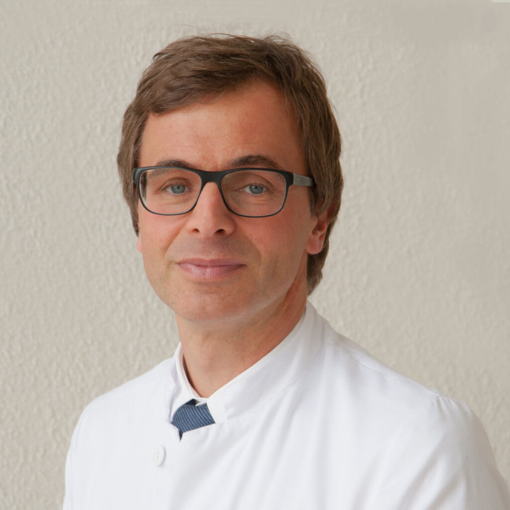
Speaker:

Prof. Dr. Stefan Mattheis

MHBA, Vice-Director of the ENT Department, University Hospital Essen, Germany

Video Transcript

Jana: Welcome to our first ENT webinar in 2021. I hope all of you enjoyed Christmas and had a wonderful time with your family and a healthy start into this new year. My name is Jana Neider and I’m live from my home office today and it’s a pleasure for me to have you all online.

Before I introduce you to our speaker, Professor Mattheis, I would like to explain a few points. Our today’s webinar will focus on the Digital ENT Operating Rooms, especially a discussion on what’s possible today, what technologies do we have, and what could be the future. The lecture will last about 30 to 45 minutes followed by a 15 minutes question and answer session. Questions can be only submitted through the online chat function and will be selected and collected by myself to be addressed to the speaker after the webinar is finished.

This webinar is live and it will be recorded to be watched again at any convenient time. After the webinar, you will receive a link once the recording is available. For further questions, feel free to use the online chat function.

Now onto our speaker Professor Mattheis. Thank you for being here, Professor Mattheis, it’s a pleasure to have you there. Professor Mattheis, I would like to provide the attendees with a few points of view. So Professor Mattheis studied medicine in Bochum and worked afterwards in Bochum [inaudible 00:01:29] and also continued his education in Paris. And now he is the vice-chairman of the University Hospital in Düsseldorf. And he also owns there a profession in endoscopic head and neck as well as robotic surgery. That makes him the ideal candidate to talk about the future of ENT operating rooms. And I’m looking very much forward to this exciting lecture. Professor Mattheis, the virtual stage is now yours.

Prof. Mattheis: Thank you very much, Jana. Okay, for this nice introduction. Just one short correction, it’s Essen, not Düsseldorf.

Jana: Sorry, I mixed it up.

Prof. Mattheis: I’d like to thank Brainlab for this kind invitation. It’s really a pleasure for me to talk to you here. I am live from my clinic office. And I guess one of the major reasons why I’m here is because I was very engaged over the last maybe five up to seven years in planning and now finally realizing a complete digital OR at our university hospital, in combination with a new outpatient clinic.

And what you see right now that is the architectural drawing of this building. But in fact, it’s now open for us and we are moving right now today and tomorrow into this new building. And I would like to give you some impressions about what we had in mind and what we were able to realize also with the support of Brainlab in realizing this digital OR.

Now I would like to change again to my screen. Yes. First, I do have to tell us that we are cooperating and have been cooperating with Brainlab. I would like to express what my impressions are. So it’s not necessarily the impression Brainlab has. And in the past years, I received speaker honorary and travel grants from Brainlab, Olympus, and Medrobotics companies, we also mentioned in this presentation. Definitely, for us, it was very important to cooperate because we are users. I studied medicine, as said, I’m not really in all those IT-specific problems. So cooperation is definitely important in order to realize and to finalize such projects.

I would like to talk about the major missions of a digital OR and what we had in mind. There were four major topics which are integration of different kinds of information. It’s not only photo and video information about patients, but also diagnostics, personal data. Everything that belongs to an OR in preoperative planning, in making resources available, and in using them.

The second major part is surgical planning. Surgical planning does not only mean making an appointment, maybe taking a look at different CT scans and whatever. But it means nowadays, especially in ENT, interdisciplinary discussion about what is to do, what will be needed, what will be the surgical plan, and how can we get support via different devices, not only navigation, in order to be successful and in order to achieve our surgical goals.

Documentation is not only for legal aspects very important for us today, but also for education, for improvement of therapy, for recalling information, which we collected in this OR. And it can be tricky nowadays, and I will show some solutions we created in our OR.

The last point, this became especially important over the last maybe 12 months because communication means today what we are doing right now, communication via web-based apps, for example. And this completely changed within these COVID times. It also means education, student education, professional education via web-based apps. And this also needs some resources in your surgical OR especially also in ENT.

When we start this integration, we have different fields, one is preoperative diagnostics. I will start with it in the next slide because also this is very important nowadays. Then the integration of different devices, and what you see here is only just a shortlist of devices. It can be extended because nowadays we are somehow dependent on medical devices in our OR, which supports us, which help us to do microsurgery, minimally invasive surgery, and so on. And in the end, it leads to an intraoperative visualization, which has completely changed over the years. And I will show you what is nowadays available.

When we start in our outpatient clinic, we always have to recognize that ENT is a visually discipline. That means we connect a lot of videos and photos from findings, for example, using the microscope to document perforation of the tympanic membrane, using the endoscope to document findings in the larynx, laryngeal tumors. But very quick, we end up in video documentation like stroboscopy, like videos of vocal cord motility.

Nowadays, it’s easy to record them and to store them. But it happens usually in this unit, and then you have your video on a hard disk drive, for example. And it’s very difficult to make it available. If you would like to have a photo documentation in your OR, you could access your hospital information system. But usually, the amount of [inaudible 00:09:01] documentation is limited to a few megabytes because hospital information systems are not made for making photos and videos available.

So what are we doing today is in many cases that we are describing our findings. That means we are describing our perforation, we are describing our vocal cord motility. It would be much easier and much more accurate to have this video documentation just in time available in your OR in order to use it, for example, to achieve correct margins in your T1 laryngeal carcinoma.

And this means already at this stage, we would like to collect all the data, put it to a server, and make it available at any place inside or outside the hospital if we would like to take a look at it. The same applies for photo documentation. Nowadays, and over the last few years, we very often took our hard copies, for example, of septorhinoplasty patients or if you have dermatological tumors to resect and took our hard copies in the OR, which is very time-consuming. And here applies the same, doing the photo documentation in your studio, collecting the data, storing it, putting it on a server, and making it available in your digital OR.

It is not limited to photo or video documentation, but also all the other diagnostics we are doing. For example, threshold audiometric measures, if you’re doing septorhinoplasties, you would like to have them in the OR available, not as a hard copy because changing to electronic chart means you also have to make this available in all phases electronically. And this also means that your digital OR has to have access to this [inaudible 00:11:26].

And if we look at all the devices, the instruments, the implants, and whatever we are using in the OR, we have to know prior to surgery whether they are available. If you are doing a stapesplasty, you need different kinds and lengths of prostheses available and you have to be sure that they are available in your OR. You can count them and maybe document it somewhere. But much easier and much more secure is regular scanning of all disposables for implants or medications when they are entering the OR.

This is a picture of our sterile floor in the new building. And when disposables arrive, and sterile goods arrive, they are scanned by the nurses directly before they are put into the storage. And you definitely are aware whether you are equipped with the necessary equipment to complete your surgeries. Showing this picture allows me to give you another aspect. While planning this OR, for us it was very important to create comfortable workplaces for all members of the staff. And this is the workplace where the nurses prepare their sterile OR tables. This is where the storage of the sterile goods takes place and around the corner is a larger storage.

And what is possible is to take a look outside to see what’s going on on the streets. Maybe if the weather is changing, which could be necessary if you’re arriving with your bicycle to work. And what we see here is a window to the OR. So even if you’re working inside the OR, it doesn’t necessarily mean that you have to go into the sterile floor and I’m not able to see daylight over the day, especially if you’re doing longer surgeries. So digital OR in more extended meaning means also creating comfortable workplaces in which you really like to work and which really supports your work over the day.

Let’s come to intraoperative visualization. What you see here is the bus system, which is able to manage all camera devices. So all visual devices that enter in your OR can be managed via this bus system. Clearly, we do not see the patient and not the staff, it’s just the OR, but what you also do not see is cabling. Because we are now able to avoid cabling through the room using such ceiling units. And the cabling goes through these units behind the wall glass fiber cabling.

So if you look at this video unit, it’s a 4K endoscopic video unit, you can place it wherever you would like to in the OR, which is sometimes necessary if you have left-handed or right-handed surgeons. If you maybe combine different approaches, if you have different teams, this could lead to a different position of your device. In fact, it doesn’t matter because the cabling is always through the ceiling unit. And you can place it on each side of the patient’s as demanded.

The same applies for this operating monitor. This screen is made for the surgeon, it’s a 4K 3D 55-inch screen. And it can be placed at each side of the patient, and again, all the cabling is invisible. So you are free to move around the patient. Using 4K endoscopes and using 4K 3D monitors means the amount of data is really high. Recording the surgery using 4K endoscopes creates gigas of storage and also the bandwidth has to be high in order to avoid lagging. That means we are using glass fiber cables and you need a separate server station in order to store the incoming video data. And then you have to process it, you have to edit it.

You can already do it immediately after surgery on this bus screen. You can do it later on from any working station in your clinic. But important is, if you have edited it, if you have maybe shortened the length, you can send it to long-term storage where it is available in the future. So if you maybe do a revision surgery or if you do control endoscopies, you can easily open up your original surgical files and can compare them to the endoscopic finding in the next surgery.

This is a closer look at these ceiling supply units. And what you see is that you can plug in with a simple solution, all different kinds of sources. It could be a microscope, it could be an endoscopic camera system, it could be a macroscopic camera system, you could use ultrasounds, and so on. So all kinds of different devices using the OR can be plugged in these ceiling supply units, you see those different adapters. And that leads all of the information to this central bus monitor where you can manage it, where you can send your picture to a different monitor and can supply, for example, the nurses with information during surgery.

The endoscopic camera resolution has developed over the years, and you can see after each invention of higher resolutions, also the surgical strategies changed, especially after 2007 with full HD cameras available. The surgical procedure changed from using one endoscope in front of your eye, which means you didn’t have a binocular view here. This resolution wasn’t high enough to do surgery by a monitor. But using full HD means the resolution was acceptable for identifying different relevant anatomic structures. And this led to a shift towards surgery via a monitor.

Nowadays using 4K and in the developments, we are now already available our 4K 3D camera systems. Again, the resolution is much higher, which gives you much more information during surgery, which allows you to have larger screens. But in the end, the amount of data you are creating this, again, much more than using full HD camera systems. And this leads to completely different structures because loading the surgery on a hard disk drive with a 4K 3D resolution means, in some cases, you have download times of hours, which has to be done overnight, and somebody has to take care about.

Another effect is that the screen becomes larger, like home cinema. Nowadays, we are using 55-inch screens in the OR, this gives you a much more accurate view of your anatomical structures. Due to the higher resolution, you can place your screen closer to your surgical field, maybe 1.5 meters. The reason is, if you do the same with HD screens, you will lose your accuracy. You will see the single pixels, your picture will get pixels, and that means the distance has to be larger. Using 4K means closer to the surgical field, higher resolution, much more detailed atomic structures.

The other thing is that usually nowadays we are sitting, you can place the monitor at the height of your eyes, which is very important because if you constantly have to look up, you feel definitely uncomfortable. Human beings normally have a look down 90% of the time over the day, and that is comfortable. So having seen the screen on eye level is very important. And in combination with sitting, it again makes you feel comfortable during surgery, which becomes important if you are doing, for example, a three or four-hour skull base surgery endoscopically. You still have to be relaxed after three hours of surgery.

Here, again, we see the screen, 55 inches, and you can place it at any height you would like to place it. You can turn it around, you can move it around the patient. It’s personally different where you place it more to the head of the patient. We usually do surgeries directly in front of the screen, which leads to the fact that you do not have to turn your upper body. And in the end, it makes you feel more comfortable.

All the features of the OR allow you also to create different atmospheres and using, for example, blue light makes it more comfortable if you are looking on the screen. There’s red endoscopic videos. Because the contrast between blue and red is better than using white light. And it’s even better if you’re using green light. So, sometimes it helps if you are cooperating with ophthalmology department. And this, for example, could be a scenery for an endoscopic skull base surgery.

So now if we look at different medical devices, we are using nowadays regularly, this picture shows you the dilemma we are in. This is a procedure in interventional radiologic OR, it’s already an integrated OR. But we brought our robotic system. We have this [inaudible 00:24:29] here, it was an intervention in vascular malformation where we did a [inaudible 00:24:36] therapy transorally. And using all those different components leads to a very crowded area around the patient leaving not enough space to do comfortable surgeries.

One example, for example, is using robotic arms, which started maybe two years ago or so into surgery. Initially, robotic arms were used in neurosurgery. We use this arm in the first step as an endoscope holder. And this is a setting from our old OR. And again, the difference is all the cabling necessary, that’s the anesthesiological device, but also for our surgery, which now completely is behind the ceiling. What we saw here is an endoscopic orbital surgery and we used this program of the robots as an endoscope holder in order to get a second free hand to do and to perform the surgery.

Over the last 10 years, we experienced robotic devices for transoral resections of malignant tumors, starting with the da Vinci system. You can see the setting. And again, also here nowadays, there is the possibility to give the signal to different kinds of monitors. Not necessarily mounted to the surgical unit, but you can place them in the wall, so making information available for the complete team, not only the surgeon but also the nurses and maybe potential visitors for educational purposes.

The signal can be routed to all different places. And also the cabling here between patient and the unit is not really necessary. Knowing that we have different devices means plugging these into the ceiling units and then using the infrastructure behind the wall in order to move resources.

The last step in robotic surgery nowadays is flexible robotic surgery. There are different systems available. This is the da Vinci flexible robot, which has been experienced over the last two years. What we were using is the Medrobotics flexible robotic system. And again, it is an endoscopic device using an HD camera system on the top, that means the signal is a 3D camera system. The signal goes into the robot, all the information also leaves the robot and comes in the robot digitally.

And when starting the surgery, we also needed an additional screen, it’s a 3D HD screen. But nowadays it’s possible to route the signal from the robotic device via the robot to all the different monitors. And in the end, also, we can communicate. I will show some examples in one of the next slides via internet with other clinics, with other [inaudible 00:28:54], and so on. Again, the amount of data is a lot because it’s a 3D signal, which means two times HD, and imagine reaching the 4K 3D camera systems [inaudible 00:29:12], again, becomes more.

Here we see an example of endoscopic surgery. And not only for educational purposes, it’s quite important to documents these surgeries. Because different steps like ligating the superior laryngeal artery in this situation and maintaining the surgical margin are important maybe to recall, because in case of recurrence or in cases of bleeding, you could, if you are able to store this and maybe using this shorter clip, as a surgeon, you can recall all this information immediately prior to your revision surgery, for example.

Especially in controlling tumors over the years and follow-up screenings, this is very helpful information after having performed the surgery and then doing control endoscopies in those patients. But also, if you would have bleeding maybe due to clip slipping in this situation, you could exactly see where you have placed the clips.

I already mentioned it, nowadays available are also for 4K 3D systems in external camera systems, which also provide additional information for a surgeon and can help you in certain situations to improve your search results. Here we see an external [inaudible 00:31:08] surgery.

And this is a device which might substitute microscopes in certain situations. It gives you an open surgical field. The complete team is able to follow your surgical steps. The surgeon himself has the possibility to switch between a macroscopic external view and some sort of microscopic view using this external camera, which can be placed at different positions in your surgical room. This information also goes through the robot, this is a robotic arm, and then can be plugged in, can be managed by the bus system.

And you can route this surgical video to all the different monitors, you can also route it inside your building and you can route it outside of your building. So if you would like to do it in educational purposes, you can send it out.

The next thing is surgical planning. This webinar is not really dealing with navigation systems, but it’s definitely one part of our important surgical steps. So, what we always have available are slices and different views of tumors. Here we see a larger and [inaudible 00:32:42] tumor invading the orbit, reaching the skull base. Definitely, you would like to resect such tumors interdisciplinary, and you have to talk about strategies. Strategies are very much depending on the histopathological findings of such tumors and definitely are depending on how much these tumors are infiltrating vital structures or functional structures.

And what you are able to do nowadays is to segment such a tumor from different anatomical structures in order to create a three-dimensional aspect of this tumor. And, sorry, I just have to plug it. In order to create a three-dimensional aspect of this tumor, and to do exact surgical planning. Exact surgical planning means that prior to surgery, definitely has to be clear which part of the anatomy has to be resected, which disciplines have to be part of this surgery, and it definitely goes further. I will show you some examples.

The possibilities we nowadays have is that we can also auto segment relevant anatomic structures like, for example, the optic nerve, chiasma, or structures in the rhino bases. So we can bring together this anatomical information with the segmentation of the tumor, and this helps you to create a surgical strategy prior to surgery usually interdisciplinary. And if we now look at the result of such a planning, we see that you get a very exact and detailed information about how the tumor is influencing different anatomical structures. And you can define critical areas prior to surgery and can discuss the strategy in, for example, this area of the orbit.

All this information and this pre-planning takes place maybe a day prior to surgery. If you make this available and send it to your digital OR and doing the surgery, you will be able to use this information in your team. What you could also do is highlight, for example, those areas where you think it could be critical. And then you can control during surgery whether you have reached this area or not using your navigation system. If you check instruments, you can also follow the route of your instruments, and after surgery, recall whether you have reached this area or not.

And there is another step. Over the last maybe five, six years radiomics became more important. And that means that not only it started in neurosurgery but also head and neck surgery data is available. It means that you not only look at the borders of the tumor, in such a case you have a histopathological finding, but what you do not know is where you find activity, where maybe after radiation where are still vital tumor parts which have to be resected.

Nowadays, especially in sarcoma surgery, we are having a multidisciplinary approach so patients are receiving chemotherapy, later on, you get a local therapy. And this consists of surgery and/or radiotherapy. And it might be important to identify vital tumor parts in order to be very successful during surgery.

And that means…so this is, for example, the study for [inaudible00:37:20.919] head and neck tumors, you see some ENT surgeons have been part of the study. That means by looking at the radiological image of the tumor, you can do phenotyping, which might help you to identify aggressive parts of the tumor. They might maybe choose a larger resection margin. And on the other hand, you could maybe identify parts of the tumor, especially if they are reaching vital anatomy like the carotid artery. You can maybe estimate whether it’s helpful to leave tumors in this area.

In combination with robotic surgery, and this is a publication from 1988, the first use was in 1985. You could use this precise and accurate information in order to do a minimally invasive surgery. So biopsies have been taken already in this area, maybe not in this setting, it was meant to be very sterile. But this has been a robot, it was an industrial robot, which was fitted with CT scan data, three-dimensional data, and then brain biopsies have been taken.

So coming back to our patients model here means if I identify a very aggressive tumor part in this area, so this is something which will happen in the future, I could maybe increase my resection margin, which would maybe mean that we would have to resect parts of the orbit, doing exaggeration. And on the other hand, if I identify less aggressive parts, we could maybe change the surgical strategy.

And using this technique in digital OR means nowadays that we can use mixed reality. So using the Magic Leap system, for example, enables you to project such surgical planning into a virtual room. And if you have interdisciplinary tumor conferences, it will enable every member of the team to follow all these strategical planning prior to surgery.

Another implication could be that you explain your surgical strategy, for example, to the patient. So here we see neurosurgical planning, but also in ENT and maxillofacial surgery, it could be important to explain to the patient where’s the tracks of the surgery are. And maybe if you would like to involve them in your surgical strategy and inclusive decisions, it could help to create an awareness about the tumor.

Here, again, we see this algorithm, many groups are working on it. At our university hospital, we have different departments, which are only working on big data and artificial intelligence in order to improve this algorithm. So we get an image of a tumor, we have a histopathological finding. This is processed. We are including clinical data, which means not only histopathological finding, but also in what condition is the patient, are there different other problems which might influence the success. And then the therapy is done. And that could mean chemotherapy, radiotherapy, or surgery.

And after having advocated this therapy, there will be a re-evaluation. And what happens is that we can learn about what we do. So when we are using this algorithm, we are in need for surgical data. And surgical data is only available nowadays via your operation report. And that is usually not enough because what we would like to know is how much is resected, and did we really reach all those relevant parts?

And using navigation systems, instruments, tagging, postoperative CT scans will help us to improve the information about what kind of surgery has been done. And also in combination with this video recording, we get much more accurate information about the surgical procedures. And this will be necessary in order to be part of this algorithm and in order to achieve statistic learning.

Documentation does not only mean diagnostics like in this case, but it starts with it. So having the possibility to use, for example, narrow band imaging for evaluating a vocal cord tumor gives you much more information than describing, for example, a T1 tumor with an extension in the anterior commissure.

What we see here is that probably the extension is a bit more than just the anterior commissure, maybe it’s reaching the right vocal cord. And this would lead to a different kind of treatment. What we also see is a deeper infiltration in the posterior part, so maybe we could be in doubt whether it’s really a T1 or maybe it’s a T2 tumor in this situation. Very important is to do this diagnostics and the other thing is to store it, to make it available, and to have it available prior to your surgical therapy.

Same patient, 4K 3D view of your vocal cord and now using blue light. And again, what we see is a much clearer margin of your tumor in the anterior commissure, extending to the right vocal cord, and also a much clearer margin to the lateral part. So using this documentation in the OR in comparison to a description of your tumor enables you as a surgeon to be more accurate in your surgical therapy,

If you are doing larger reconstructions and would like to reconstruct or have to reconstruct your pharynx, for example, there’s a free flap, then one question is always, how patent is my anastomy? And also here we can use video documentation with ICG videoangiography. So we are giving ICG intravenous. And some seconds later, we see the profusion of the arterial and venous anastomosis. So what you could clearly see is, is there stenosis, is it a patent anastomosis, you can count the time. And this gives you much more information in case of problems. You can recall what you did, and again, it means video documentation of surgical results.

We had a cooperation with our Robotic Department at University of Duisburg-Essen with Professor Kecskemethy. And we tried to analyze what happens during our robotic endoscopies. And that means we matched the endoscopic view to a model, which was created after using CT scans and MRIs. And the aim was to establish a model or an algorithm which helps us to evaluate whether special anatomical parts or tumor parts are reachable with our existing endoscopes and instruments. This is just the role model for all different surgical evaluations we would like to do.

So in the future, if we are able to record what we are doing and maybe are able to receive data to create a model, we will much more be able to recall what we did during surgery. And this will help us to improve with our innovations regarding surgical devices, regarding camera systems, regarding robotic devices, and so on.

This is another example when we started using a robotic arm in clinical trials, it was also… QVC is this robotic arm used as an endoscope holder in endoscopic middle ear surgery. It is important to evaluate in what position we get the best results. And therefore we need to collect this data from the surgical robot. And here we see how the robot moves the endoscope during surgery. And the combination of robotic data and endoscopic data helps us to evaluate where we can improve these surgical results.

Last point was communication, and I already mentioned it. Nowadays we are definitely using more and more transmissions for students’ education but also for professional education. This is a picture of the ENT German Congress in 2019. And we did a live surgery transmission from Essen 600 kilometers to Berlin using surgical robots to resect a [inaudible 00:49:20] tumor.

This is an endoscopic surgery, we had a 3D HD view, transmitted was this HD view of the surgery. And there was a constant interaction possible. That means we had an audio connection, we had video connection, questions could be asked during surgery. And at the same time, we had a panel in Berlin able to communicate with the surgeon. And we see on the right, this was a cooperation with a clinic from Pittsburgh [inaudible00:50:05.457]. And we were able to perform this surgery and to communicate with the audience.

Another example is the Live-Surgery broadcast in cooperation with the European Laryngological Society. And what we see is over the past five years, it has been established. And unfortunately in 2020, we were not able to perform this last surgery event due to corona because it would have made sense to do it online. But we were not able to do the surgeries with different surgeons at that time, in 2020.

But in 2019, we had 35,000 viewers from nearly 100 different countries. And imagine this difference, not only having maybe 3 viewers and visitors in your OR looking over your shoulder, but 35,000 people watching your surgery, listening to the surgical strategies, and maybe learning how different clinics do different surgeries. And afterwards being able to be part of the discussion if you have questions and answer them.

This needs a really complex infrastructure because you need the camera system, you need the transmission system, you need to connect your OR to the internet. And then you have to have a stable connection in order to get a good quality. But integrated ORs nowadays are able to provide all these infrastructures and make it possible to do such transmissions into the student auditorium, for example, you can send it to your assistance in order to educate them. Or you can take part in such international global live surgery events in order to transmit it in different countries.

To conclude, this is the red light, which is not used during surgery, only after surgery. In a digital OR, we are achieving an improvement in integration of all those complex devices we are using nowadays. We have much better opportunities to do surgical planning. Documentation is easier and it allows us to do communication… With every place we would like to communicate with, in the end, also, we could send it into the internet.

Future aspects are using this infrastructure in order to collect a lot of data in order to learn what we are doing, to learn how we can change our surgical strategies. And making it possible to get additional help in our decisions using artificial intelligence not only for the interpretation of CT or MRI scans, but also in order to interpret our surgical strategies. Thank you very much for your attention.

[00:53:54]

[silence]

[00:54:14]

Jana: Professor Mattheis, thank you for the interesting insights on your new OR that you have built in Essen. I’m sorry for mentioning Düsseldorf. It’s because I’m always flying to Düsseldorf and now I mix that up. So of course, it’s in Essen that I always go to, and I also saw that installation live. And it’s really interesting to see how much technology you have been able to implement in these ORs. And I’m also sure that you’re gonna reach your personal goal to become paperless within your OR. So I see that you go step by step to that aim and all the technologies you presented will, of course, help you with that.

And I believe it’s very important so that clinicians and the technology companies working closely together because you have all the clinical knowledge and we need to implement that and find out how to really digitize your surgery. And make sure that all the data that you create and evaluate during the surgery is also being backed up and stored somewhere. And then also made sure that these can be used afterwards. And I think you presented that very nicely.

And we also received a lot of different questions because you also had a lot of different topics from a technology perspective. So I would like to address them in a second. But I also would like to remind everyone, you can still use the chat function to send further questions and I will still address them. Okay, Professor Mattheis, the first question to you is a more technical one. And the question is, is the server locally hosted or do you make use of cloud services?

Prof. Mattheis: It’s locally hosted. I’m a user so I’m not really maybe the right address for all the technical aspects. But we had to establish, and establish an owned server just for this OR and the outpatient clinic receiving all the picture and video data. So it’s locally served server established just for this OR.

Jana: Thank you. Yes. So the installation, Essen is completely on a locally hosted server, and all the different technologies that have been shown, like the endoscope video signals, the navigation system, and so on, they are all combinated with each other and can be then accessed through the bus system that Professor Matthies showed.

Then a second question, and I think that question arised before you showed the mixed reality. But the question was, there’s a big push for augmented reality headsets to replace screens in the OR, what is your opinion of making use of these devices?

Prof. Mattheis: So, right now they are not exactly enough. And so there is definitely a difficulty to match your augmented reality using those goggles with the reality. So we are talking about mixed reality. What you can do is nowadays, already you can augment, for example, your navigation system views, you can augment your endoscopic screen, for example, by pre-planning and marking relevant anatomic structures. Definitely, the accuracy of all these systems is still an issue because imagine, if you’re having tumor extension to the optic nerve or to the carotid artery, it really depends on millimeters.

So even if we are able to use all those technologies in order to support our surgical procedures, it’s still more relevant to identify anatomical structures maybe via a high video resolution, and then maybe to match them with the augmented information. But it is only a matter of technology. And what we saw over the last 10 years, for example, is that the accuracy of navigation systems, and also the accuracy of using augmented reality very much improved over the last few years. And this will be the future. Right now definitely it’s not yet possible. What you can do is preoperative planning and intraoperative support, but not using it straight during surgeries.

Jana: Thank you. I totally agree with that. So I think the industry is working heavily on that and being able to do pre-planning and patient education as well as teaching students or even having a final check of the data set before the surgery starts live in the OR, I think, this is already adding benefit. But, of course, the aim is to do real intraoperative navigation and then really get rid of the screen using mixed reality.

The next question is, actually, after having one year of a corona pandemic, and you also mentioned that quite a lot, what options do you see to improve our daily ENT work with the technologies that you just presented? So is there anything specifically that you could recommend to a colleague that he could use to now even improve and… I mean, I have a personal take on that, but I will let you answer first.

Prof. Mattheis: I’m not quite sure whether the impact of corona…let’s say difference. What happened is that definitely surgical strategies changed due to corona. For example, the indication for larger surgeries sometimes were changed, especially in Great Britain, [inaudible 01:00:20], and Italy, in tumor surgeries, the centers tried to avoid, in some cases, larger open surgeries, and it shifted a bit towards endoscopic, more or less minimally invasive surgeries. Nobody knows whether this was a good idea or not because we do not have any follow-up results of all those therapies.

Definitely, everything that has to do with education is much easier doing it with a transmission with online resources. I’m not quite sure whether changes in the daily practice are dependent on those technologies if they are or have been caused by corona. And definitely, we need more evidence, what really happens to the patients if we change therapeutic strategies. So I think for educational purposes, it’s really important, especially what we’re doing here right now is the best example for it.

Jana: Thank you. I totally agree with that opinion. And I also see that they, of course, could be done more. But having the opportunity utilizing the technology you have to stream also information directly to the internet or to a larger room where the students can sit is definitely a benefit. And also using the mixed reality technology now to really educate students without touching a patient, but them still seeing the patient live in front of you, I think, this is really adding a lot of value. And I will also contact the person who sent that question so we can also follow up on that.

The next question is, so you showed a few instruments that have been tracked under navigation, and the question was, was that being tracked with electromagnetic systems or with an optical system?

Prof. Mattheis: So we have both available and are using both. And it has…both of those have advantages and disadvantages. So the tracking itself has a different purpose. It really depends on where you are using your system. In general, we see a shift a bit more in ENT surgery towards electromagnetic systems. But especially if you’re doing interdisciplinary surgery, many neurosurgeons are definitely using optical systems. So it really depends.

Jana: Thank you for answering to that. So, of course, you always have the option of using optical as well as electromagnetic systems. And I will also follow up in a second with our next webinar that we’re gonna advertise because this will especially focus also on that question. But before coming to an end, I have one last question and I think you answered that already. So the question was, are you also documenting or streaming all of the data to students? You answered quite intense in regards to the students for the streaming, how do you see documenting with the technology?

Prof. Mattheis: I think there is a definite need to document at least relevant parts of a surgery. What we see, especially in the United States, is that you could as a routine make video documentations of standardized procedures, for example. And there are systems available already and software modules available where your surgery is rated. You could also achieve a peer review rating of your surgery which could help especially younger colleagues to improve their surgical techniques.

This applies especially for standardized smaller interventions. If you have smaller surgeries where you have definitely always the same steps. And if you document them, and then maybe review them together with an experienced surgeon, it definitely helps you much more. The other thing is you could also define goals prior to surgery. And then definitely try to reach those goals, document them, and later on, if you review it, get information whether you reached your surgical goals or not.

If you have standardized practices like suturing, like applicating certain devices, you could also review them, because you could just measure how long did it take to complete this surgical step, for example, and then you can compare it. So this is already available, it’s already in use. And there are educational programs especially in the U.S. which are focusing on this, for example, in transoral robotic surgery because it’s very standardized. And you could define definitely those surgical steps prior to surgery. So I…

Jana: Thank you.

Prof. Matthies: …strongly believe that you get a lot of information not only using maybe big data but also just reviewing your own work, you get so much information by documenting as a routine your surgical results. Our aim is…so we are just starting with this. But our aim is that we get documentation of all relevant surgical steps and systems available which make it possible. So using just maybe one button in order to start a video and stop it from your surgical field will allow you to do so. If it is very complicated and if you do not have the storage, if you do not have the devices that somebody has to take care about it, you won’t do it. But in the future it’s much easier.

Jana: Thank you for all of this insight, Professor Mattheis, and thank you for this fruitful discussion. That was also the last question we had. And I think it was really interesting to talk with you about the future of ENT operating rooms. Thank you also, everyone, who joined today. I hope you really liked the webinar.

And I would like to also guide you to our webpage where we have a lot of information on digital operating rooms so you can have a look at that online, where you also find detailed information on how integration, documentation, and communication especially work. Because I think there was a lot of information in that webinar, so here you can have a follow-up. We also have some customer stories here at that webpage that will help you to find the right solution for your OR since this is always, of course, scalable.

And then I would like to thank you, Professor Mattheis, and highlight that the next webinar is on 24th of February together with Professor Stelter. So today we saw that there was also few questions on navigation, so we’re gonna have another webinar on navigation and functional endoscopic sinus surgery and skull base surgery. And we will really focus on why, how, who, and when to use it. And I think this is all important questions, and Professors Stelter will be happy to answer all of that.

If you are now curious on more of our webinars and news from Brainlab, please follow us on all social media channels or drop us an email to [email protected]. Thanks again, Professor Mattheis. It was a pleasure to have you here today. And thanks, everyone…

Prof. Mattheis: Thank you, everyone. Thank you, everybody, for listening.

Jana: Thank you for joining, everyone. Stay healthy and goodbye.

Prof. Mattheis: Bye-bye.

Related Webinars

mixed-reality
Digital O.R.Digitalization+ 1
Talking Innovation – Today’s technology and what’s coming in the future

Technology continues to rapidly evolve. With this, we ask: How is today’s technology impacting healthcare? And how do we see innovation shaping its future? Join our speakers as they share not only their answers to these questions, but their expectations …

Simon Enzinger
Woosik Chung
Jennifer Esposito
Auke Meppelink
digital-operating-room
Digital O.R.Digitalization+ 1
Building a Digital O.R. Roundtable Discussion

Creating the perfect operating room with the latest technology that suits the needs of different departments is a complex task. During our discussion, you’ll hear the opinions and experiences of various key roles involved in the process of building a …

Stefan Mattheis
Marcello Bonfim
Thomas Fritsch
David Krusch
Philipp Wolf
digital-o.r
Digital O.R.Digitalization+ 1
The Benefits of a Digital O.R. in Daily Practice

Digitization in hospitals is a topic of growing importance, especially in the O.R. In this session, we will bring together different perspectives from around the world to share the advantages of having a digital O.R. and how it has changed …

Xavier Escayola
Albert Busch
Daniel Köbsch

See more upcoming webinars

Register now