Are you in %country%? Do you want to visit our website in another language?

Webinar

Navigated Ultrasound in Neurosurgery - Ask the Experts

Topics
Image Guided Surgery
Cranial Surgery
Intraoperative Imaging
Neurosurgery
Surgery Navigation
Language

English

Share this page

Description

Brainlab invites you to a live webinar: “Navigated Ultrasound in Neurosurgery” on 17th September at 5:00 PM (AEST, GMT+10) hosted by Dr. Awad and Dr. Bartos, both experienced ultrasound users. Join their discussion around the applications of navigated ultrasound in neurosurgery, where they sharing their learning curve to fast track yours and answer your clinical application questions.

This webinar will cover topics, including:

– How to best start using ultrasound in neurosurgery?
– How to interpret intraoperative ultrasound images during the course of tumor resection?
– What to consider before and during surgery to optimize ultrasound usage?
– Q&A session with the audience.

We look forward to meeting you online!

Language | English

In case you can not join the webinar, it will be recorded and shared afterwards.

Speaker:

Mohammed Awad, MD
Mohammed Awad, MD

Neurosurgeon, The Royal Melbourne Hospital Australia

Michael Bartos, MD
Michael Bartos, MD

University Hospital Hradec Kralove Czech Republic

Video Transcript

Uli: Good morning, good afternoon, and good evening. Welcome to today’s session on navigated ultrasound for neurosurgery. First of all, thank you for taking the time to join us. My name is Uli. I work for Brainlab, and I will be your host for today. It is my pleasure to introduce Dr. Awad and Dr. Bartos as our presenters, experts for today’s session. They will share their experience using ultrasound intraoperatively and will happily answer all your questions. Dr. Awad and Dr. Bartos right now are the perfect role model for social/physical distancing because they’re being on the opposite sides of the globe. But what brings them together is the passion for ultrasound.

Dr. Awad joins us from Australia. He is a trained neurosurgeon. He trained in the UK, where he also started using ultrasounds and he’s been using ultrasounds since 2010. He moved to Australia six years ago and now works as a complex spine and neuro-oncology consultant at the Royal Melbourne Hospital. Dr. Bartos joins us from the Czech Republic. He’s a consultant neurosurgeon at the University Hospital of Hradec Kralove. His main interests are neuro-oncology, functionally-guided resections, and of course navigated ultrasound. He’s been a navigated ultrasound user since 2013. He’s a frequent international guest speaker and a Brainlab R&D consultant. His research interest is also around developing a perfect ultrasound phantom for training.

Our agenda today will be that Dr. Awad will start with a presentation, introduce navigated ultrasound, and talk about his recent cases. Dr. Bartos will follow up with some more of his cases, give more tips and tricks. Both presentations will be around 15 minutes, which will leave us enough time within this hour to do a Q&A session together with you. We have, therefore, enabled an ask-the-question feature in your interface, where you can pop in up a question at any time and we will make sure that we address the question. We want to collect the questions not to interrupt the presentations too much so we have enough material for the question and answer later on.

We have also prepared three polling questions to get your collective feedback, which will help us further to tailor our discussions. And if you miss anything, don’t worry, we will be sending around the on-demand recording when it’s available. If you just joined, again, welcome to our today’s session about navigated ultrasound with Dr. Awad and Dr. Bartos, both very experienced ultrasound users. And with that, I’d like to hand over to Dr. Awad.

Dr. Awad: Thank you, Uli. Let me share my screen. Hopefully, you’re all seeing that at the moment. So, thank you, Uli, for that lovely introduction. My name is Mohammed Awad. I’m a registered neurosurgeon currently working in Australia. And I’m going to give you just a very quick flavor of intraoperative navigated ultrasound use. I’ve been doing this for about 10 years now. And I’m going to try and share some of those nuances with you today. And like I said, this is just a flavor in 15 minutes for those of you who haven’t used ultrasound before, and then Dr. Michael is going to enhance on that, go over some of the same aspects that I will talk about and also enhance on that and maybe go into bit more detail. It’s going to be an interactive talk so we’re both going to interrupt each other’s talks and make it interactive and sort of a bit more entertaining for you as well and trying to keep you all awake, especially for those of you who it may be at the middle of the nightfall.

So we’ll start by the first polling question. Now, you should get a little screen coming up on your computers at the moment. And we just want to get a flavor for those of you who are using ultrasound at the moment versus those of you who just may be using standard navigation, those who are using navigated ultrasound, or those of you who are not using anything at all. So, for those of you who are, indeed, cranial neurosurgeons, please answer this question, and we’ll give you 10 or 15 seconds to do that and then we’ll share the answers. And it’ll be interesting to see from all of us around the world here what we’re actually doing at the moment and how we’re operating because I do understand that we literally have people participating from all over the world.

So why don’t we look at some of those answers at the moment? We’re not getting any answers at the moment. It’s coming up at 0%, so I think that’s probably an error.

Dr. Bartos: It looks like a technical problem, I think.

Uli: I’m seeing navigation standalone 41% of the users have answered, ultrasound standalone, 17% of the users, navigated ultrasound 30%, 3% don’t use any ultrasound, and 8% don’t use navigation.

Dr. Awad: Well, that’s interesting. So there’s 30% of people on here who are using navigated ultrasound already. So a lot of you may have a lot more experience than me or Dr. Michael. But really I’m going to try and keep my talk for those who have not used it before, and Michael, like I say, will enhance on that a little bit more so. So ultrasound, like all the other tools we use in neurosurgery for centuries, is another tool that we use to visualize in what’s going on inside the brain. Now, we’ve been using these kind of tools to either get access to the brain or to visualize what’s going on inside the head, like I said, for centuries. Ultrasound is actually one of the first instruments of its kind that we used like this back in the 1800s, in fact, and early 1900s. And to some degree, it certainly, in certain aspects of neurosurgery, a lot of people continue to use that, for example, in pediatrics. But for multiple other reasons, we stopped using it in adult neurosurgery. And this is one of the reasons why early ultrasound pictures were like this. And, you know, when you got a picture like this, you know, we all have the same question, you know, “What in God’s name are we looking at?” and we had no idea. And so, we shied away from doing that.

And then, ultrasound became a little bit better and improved. And we started to see some form of anatomy with the probes. And we were getting pictures, where you can start to see some distinctions of sulci, gyri, various deep anatomy. But again, as neurosurgeons, we moved away from looking at these and we started to look at things like CT and MRI scans when they came about in the early ’80s and ’90s, and so forth. And so, even when you’ve got a picture like this, you know, because we weren’t trained to look at these pictures, we didn’t know what we’re looking at. And so again, we shied away from ultrasound altogether. Now, what happened was basically in the late ’80s, in the mid to late ’80s, the Scandinavians came up after Brainlab and other companies that already come up with cranial navigation, they decided that they were going to try and integrate navigation with ultrasound, and said, “Well, look, you know, with navigation, once you start your operation, your picture is no longer valid, whereas you can get these up-to-date pictures with the ultrasound and we’re going to try and integrate it.” And they came up with this machine called SonoOne. And they were, really, the market leaders in navigated ultrasound.

But, you know, there were some flaws with it and it was an early machine, and the navigation component wasn’t the best. It was actually pretty good, but it wasn’t the best. And so, of course, what Brainlab did, who have a lot of history in navigation from the ’80s, is they then decided they were going to get into this field. And they partnered with BK Medical, which is a company that has a lot of heritage in the ultrasound world, and they partnered together to make something that was better than what was available in the market before. And I’ve been using this for a few years now, and I’m going to share some of that experience hopefully with you. So what you’re looking at on the left there is a standard navigation Brainlab machine, and on the right is the bk5000, which is the most up-to-date ultrasound machine which is used for neurosurgery.

So let me talk a little bit quickly about how this works. So basically, you position your patient like you do any craniotomy. So you fix your patient and you get your start and you register your patient like you do with any navigation. The only thing you need to remember here is that when you’re positioning your patient, your craniotomy needs to be parallel to the floor so that, whenever you do come to get a picture, you have fluid or tissue interface with the probe that’s continuous and that there’s no air, as you can see with the little diagram there in the middle, because air is not your friend, it’s not the ultrasound friend, and it will give you a shadow and you won’t be able to see what you’re doing. So you need a continuous interface between your probe fluid and tissue.

On the left here, you can see, so these are some of the bk5000 probes on the left. And you can see that they’re navigators. So we put the little navigation stalls on them so that the idea is that when you’re getting a picture, it’s picked up by the Brainlab machine, and it will put it in the right place for you. So what I mean by that is, as the diagram shows in the middle there, you’re using your sound probe, and the ultrasound, what it does is it takes a 2D slice. But essentially, what it will do is it’ll take multiple 2D slices, and as it’s taking those 2D slices, the little star on top there with the balls on top will put those slices into a 3D picture and put that 3D picture into the correct place to overlap the MRI scan.

So ultimately, what you’re doing is now you’ve got an MRI scan, which is the preoperative MRI scan or CT scan that we’re using, with an updated 3D image on top that’s real-time. It’s there and then. It’s there available for you to use. And you can get that multiple times through the procedure. So this is a little video of me using it. And you can see me there doing a quarter 3D sweep. So the probe is taking multiple 2D images. And you can see in the top left corner there that 2D images are going onto the Brainlab and I will be able to now, once the computer puts the images together, navigate to that 3D picture. And it’s real-time so I can do that halfway through my resection, three-quarters of the way, at the end. Quality control, you can do it whenever you like. And it only takes about 60 seconds or so to get this 3D sweep. So you can update your navigation picture in 60 seconds. So it’s an iMRI scan, but much quicker, much less cumbersome, and it’s right there available for you and much cheaper, of course, than an iMRI scan.

So then the question is, what’s the advantage of having the ultrasound and Brainlab together? Why not have one or the other? Well, of course, the Brainlab is useful because you can guide your incision and your access as we commonly do. But then the Brainlab, in combination with the ultrasound, will reconstruct those data set slices for you as I’ve showed you into a 3D image. And then not only would it do that, it will put that 3D image into the correct place so it will overlap your MRI scanner so you’re now navigating to an up-to-date, essentially an up-to-date MRI if you like, because that’s what it’s doing for you. And you can continue to do that throughout the case. And because it’s overlapping the image for you into the correct place onto the MRI scanner, you can then interpret the image because you can see where the falx is. You can see whatever other structures that you know that you’re looking at on the MRI scan and it will put it into the correct place for you so that you can correlate it to your pre-operative images.

Of course, the use of the ultrasound is that it will do that for you. You need a probe and you need that equipment to then bring those new images onto the Brainlab. If you don’t have the ultrasound, the alternative to get up to the images is an iMRI or an ICT, which, like I say, is extremely expensive, extremely cumbersome, and it’s not as easy to use as this. And so, it provides the need to collect this update imaging and to update your roadmap.

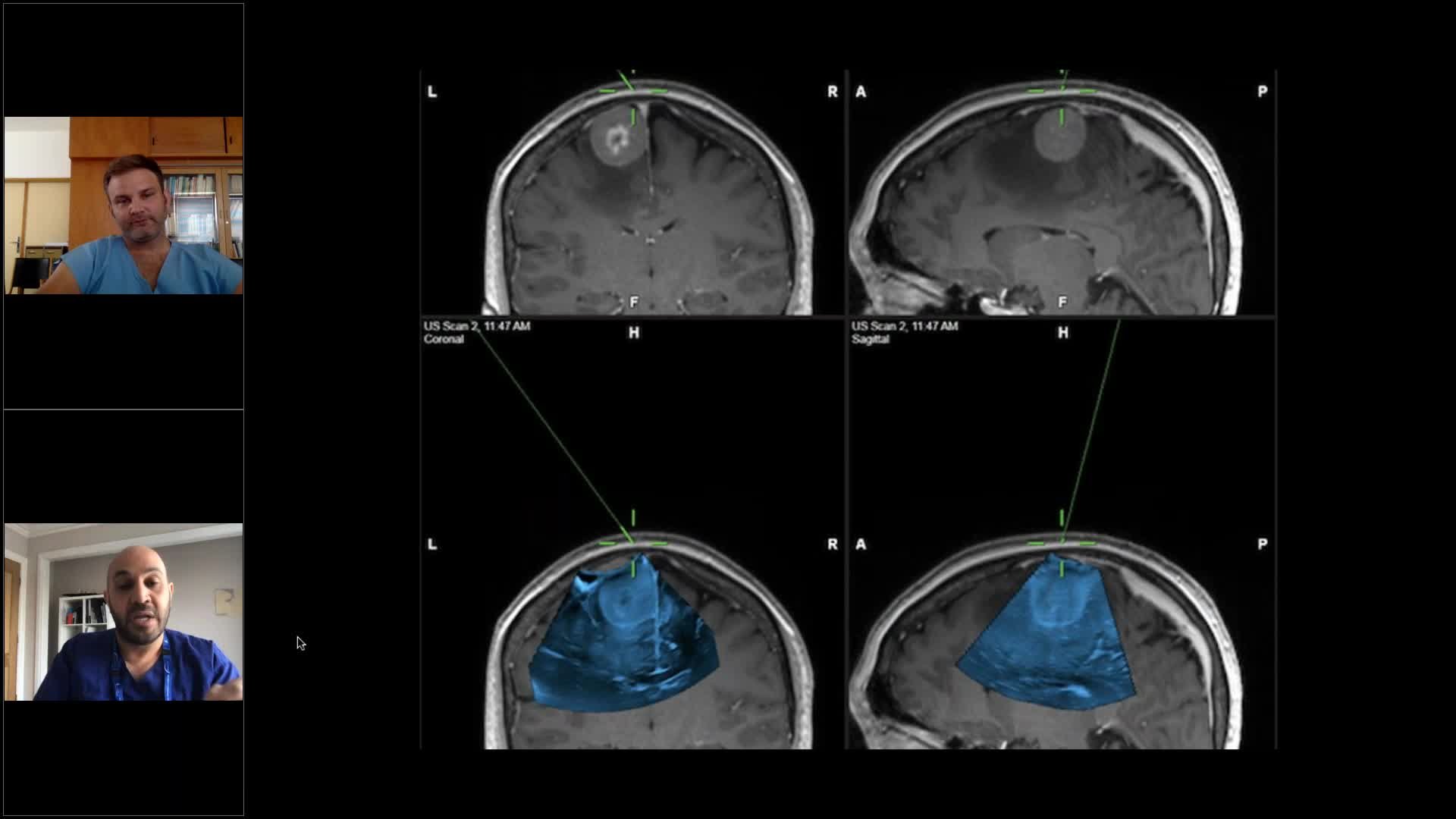
So, of course, this now begs the question, “Okay, this all sounds wonderful. You’re giving me an iMRI scanner, which is, you know, a fraction of the cost. It’s there. It’s sitting in the theater. It’s much cheaper to use. But what am I actually looking at? How good are these pictures?” Well, I want to show you some pictures very quickly. So I’m just going to show you one case, which is a metastasis. And this is a lady in her 40s with a breast metastasis. And this is the picture you’re looking at. So what you are looking at in blue at the bottom there is the 3D sweep. Now, of course, this is just a snapshot, but I can now navigate to that 3D image that you can see at the bottom there. This is just a close-up, and you can see how well you can see the falx there, and the sulci, and all the anatomy really quite well on the border setting the interface between the tumor and the brain.

And so now, when I’m putting my probe in and navigating, I am navigating to an up-to-date image so I don’t have to worry about poor image…poor registration rather and I don’t have to worry about brain shift either because this will update my roadmap as I go along. So again, just very quickly as a flavor, you can see this is a snapshot from the Brainlab screen. The top images are the axial, coronal, and sagittal slices of the preoperative MRI. The middle images are the pre-operative sweep, so with the craniotomy open and we sweep across the dura. And then the bottom image is my quality control once I’ve taken the tumor out, looking at my cavity to see if I’ve left anything behind at all. And that black cavity you’re seeing there is fluid-filled. Remember, there’s no air in an ultrasound.

And that’s just taking the blue away. You can see how it updates the images now and it tries to match it with your preoperative MRI scan. So you’re updating your roadmap quite nicely there. Very quickly, to give you another flavor with a high-grade image, you can see here this is a typical high-grade glioma. Now, most of us would resect this, and we’d resect this to the margins of the contrast-enhancing area because that’s what the evidence tells us to do. But you can see with this initial ultrasound sweep, the margins of this tumor go way beyond those two contrast-enhancing areas. And you should see a very nice interface there between more dense tumor and more sort of normal or gliotic brain around it.

And there you could see the top row is the MRI. There’s my preoperative sweep showing the tumor in the middle. And then the bottom is after I’ve resect the tumor and I thought I was pretty much done. Now, what you can see in the bottom slice set there, well, circled in red is that’s tumor residual. So you can see there’s my cavity in black, and I’ve got residual tumor there. And I can target that now and I can go back having done a 3D sweep, and I can use my navigation probe, my Brainlab navigation probe, to find where that residual is and I can go back and resect it. And you can see on the next screen, there’s my middle image showing the tumor still there, and at the bottom that tumor is now gone. It’s very important when you do all these tumor resections, again, Michael will talk to you about this in a bit, but you must try to keep your tumor cavity resections blood-free as much as possible because blood… So the two things that the ultrasound doesn’t like is air and blood. It will give you a shadow and it will create an interface and you won’t be able to see what’s going on very well. So it’s a really good way of also controlling your hemostasis as you go along. It teaches you to do that because the need is there for you to do that, of course.

This is a low-grade glioma. So just very quickly, the advantages of this is it shows up very nicely on the ultrasound. You can also get an angio sweep. So you can see in the bottom, you can see these little red dots and they’re vessels. And actually, when you’re navigating into 3D mode, those vessels become in 3D mode and you can see them and you can see where they’re running. You can see their course and you can avoid them, of course, when you’re doing your resection as I’ve done here. You can see my resections at the bottom. This is just the flavor of the angio mode. As you can see, if you get it done really well, it can show things like aneurysms and the anatomy of vessels really, really well. You can navigate around that. And it also gives you an idea of whether you’ve got flow following the aneurysm, etc. So really, really useful.

Going back to our low-grade glioma, you can see there, I’ve followed the tumor in towards the ambient cistern and I was able to get a safe, good resection because of the ultrasound more than anything else. Very, very quickly, the last case, this is just a DNET, very small lesions. And this is the advantage of having the ultrasound is, I mean, if this wasn’t in this location because most of you may just go on to do a full temporal lobectomy, but if, for example, this lesion was somewhere deep in a more eloquent area, imagine if your Brainlab or whatever navigation you’re using was off, trying to hunt for that, it’s like hunting for a grape in purple jelly. Whereas once you get your ultrasound sweep, you’re gonna get an up-to-date imaging, and you’re now navigating to a perfect location. You can go straight down to that lesion.

And you can see how well this DNET shows up on the image on the right there. You can see that hyperechogenic area which lights up as a circle there. There it is, there in blue at the bottom. And what I want to show you is this at the end. So you can see what I’ve done. So the top image is the MRI scan, the bottom image is the…sorry, the middle image is the preoperative or pre-resection ultrasound, and the bottom one is post-resection. And you can see I’ve got my probe at the back, front, top, and bottom of my resection cavity, and I can show you that I’ve taken that tumor out because the probe tells me exactly that I’ve done that.

Again, in a minute or so, other use of ultrasound, you can see this is the video showing a patient with a very large disc prolapse. And you can see how distorted and crescent-shaped the dura is then. It’s only expanded because I’ve obviously done my laminectomy above it. But you can see that big, big bubble there which is the area to the left. And you can see here now this is gone. This is resected. So you can see the dura there takes some more usual shaping. You can see that the nerve root is bouncing around in there. And this is, by turning the probe 90 degrees, what you’re looking at there is the nerves in longitudinal mode bouncing around that I know that I’ve decompressed.

So that was just a whirlwind, very quickly, of ultrasound with navigation. So what’s the uses, integrating ultrasound with navigation is a better tool I think than navigation alone because you can update your roadmap, which you can’t do with your standard navigation. It’s the same as iMRI, but much less cumbersome, of course, and much quicker, much cheaper, much more readily available, they’re much more likely to use it than having to put in a patient in and out of an MRI scanner, which takes, you know, the best part of an hour, an hour and a half each time. So you’re updating that roadmap as you go along, you’re counting for brain shift, you’re checking your resections, so there’s quality control. And also, if you get a registration error at the beginning, it doesn’t really matter so much because the ultrasound sweep will put that tumor image into the correct location and space so you’re now navigating to an up-to-date correct image. It’s not cumbersome. It takes 60 seconds to get one of those sweeps. You get really good image quality as you saw there because of the digital integration between the bk5000 and the Brainlab machine.

The ultrasound probes are also sterile. So unlike the older system, or the one I showed you before, which is the SonoOne, where you have to cover and put like a plastic sheath over the probes, these probes, they’re sterile, so you can just use them straight into the brain. And, so, of course, with experience with using this, you can get much better. And the more you look at this, the more you look at these pictures, the easier it becomes to the eye, and the more you can understand some of the nuances as well.

Now a lot of those nuances I’ve not gone through. I’ve gone through a very few of them, but Michael will go through some of them more so in a second. I’ll leave it there. But thank you very much for listening. That was, like I’ve said, a quick whirlwind on the basics of ultrasound. And before we go on to Michael’s talk, I’ll just go on to a second polling question, which I think is pertinent at this time, and Uli is going to hopefully bring that up and you’ll see that. The question is, why do you think you and/or your department is not using intraoperative ultrasound at the moment? Is it because you’ve just never been trained to interpret images is one of the reasons we spoke about? Is it just simply too expensive for your department? Or did your department have something else like an iMRI or a CT? Or do you worry about something like intraoperative infection or losing time, for example, from using it? Or you just simply never had the opportunity to use intraoperative ultrasound? So if you could answer that now and we’ll come up with the answer in a few seconds, Uli is hopefully going to either show us or tell us the answer.

Uli: Give us three more seconds. So about 30% of the audience has voted.

Dr. Awad: I think everybody else has fallen asleep.

Uli: I don’t think so. Okay. 35%. But I think the tendency is clear. Yeah, let me close it and then share the results, hopefully this time. Can you see the numbers?

Dr. Bartos: No.

Dr. Awad: No. Unfortunately not.

Uli: So the numbers are, never been trained to integrate the images, 36%, too expensive for my department, 18%, the department has other intraoperative imaging solutions like iMRI or CT, 23%, only 5% worry about intraoperative infection or losing time, and 20% never had the opportunity to try ultrasound.

Dr. Awad: That’s interesting. Well, thank you for sharing, you all. Thank you for being honest. And that gives us a bit of a flavor of what’s going on. We’re going to quickly go on to Michael now, who’s going to give us a really nice, interesting talk with some beautiful images. So, over to Michael. Thank you.

Dr. Bartos: So, thank you, Mo, for the great introduction. I’ll try and tie on to your talk and just go into a little bit more detail. In the beginning, I’ll do a little bit of cherry-picking and show you very nice images. And then later on, I’ll show you how it looks when the going gets a bit tougher and then what you need to do to get the images that you require to have good resection control. So in the beginning, I would just like to start with a little bit of a humorous note. And this is an interesting study. It’s quite old now, 1994, but it shows that our own judgments of how good we are at resecting tumors is actually very flawed. So in this trial, the surgeon at the end of the resection was questioned on whether he or she thinks the resection has been radical or not. And then following that, an immediate post-operative MRI was done. In 21% of cases, the surgeon expected a residual, whereas on the post-op MRI there was a residual in 70%. And in 71%, the surgeon expected it to be a gross total resection, whereas in reality it was only 18%. So, this really shows that our own judgment is very flawed, and we definitely need some sort of intraoperative imaging. This talk happens to be on intraoperative ultrasound. I won’t go into any much detail on the other modalities because I don’t have any personal experience with them.

So, Mo covered this very well, why image fusion? Well, I think it’s mainly because we are trying to look at specific planes, the orthogonal planes, meaning sagittal, coronal, axial. When we look at tangential planes outside of those, we tend to get lost, and integrating into navigation helps a lot with that. Not only that, but the probe itself is a narrow picture. It’s like looking into a tunnel, actually, like you’re seeing at the bottom right over there. And if you have the background pre-op MRI, you orientate yourself much better, and interpretation of the image becomes much easier.

So let’s just do a little bit of cherry-picking. I’ll show you a low-grade glioma and the SMA on the right, of course, done with neurophysiology. This is how pure ultrasound image looks in the coronal. I think anybody can interpret this very well. No problem. The sagittal, you can see the cinguli gyrus at the bottom there. When I do this resection, I always look for landmarks. If on the pre-op MRI I see a specific landmark which will guide me to where the resection needs to end, I’ll keep that in mind when I’m doing the resection control.

So this is how it looks in the fusion image. So, as you can see, if you see a broader picture, you can orientate yourself much better, the sagittal view, and then getting into the resection. So you can still see a residual towards the posterior of the resection cavity. In the front, you can see the resection cavity. This is looking in the coronal. You can turn the probe as you like during the resection, still see residual, still some minor residual, and this sulcus actually that you can see just above the cinguli gyrus, that’s the limit of my resection.

Moving on, just showing you on video as the image changes in real-time. This is unfortunately an iPhone video from the screen so it’s not great. You can see the little detrituses moving inside the cavity. I flush a lot, I irrigate a lot of saline to keep it clean and to get a better image. So I think we should just fly through this quickly. You can see the post-op. No residual. Another one, this was a complicated case, a hand-knob tumor with some functional reorganization. This is how it looks on the fusion image. You can change the color as well. This was a previous version from Brainlab, older version. You can still see the residual there. At the end, you can see it’s a radical resection.

So moving on to the high grades, it’s a little bit trickier because the high grades are very heterogeneous. You have necrosis, you have viable tumor, then you have edema, etc. It’s sometimes difficult to tell what is what. You can also use Doppler, things as landmarks, the falx. I would definitely measure the distance to the ventricle. That is a good guide, the sort of depth gauge during the resection, in this case, the sulcus below it, below the tumor. We have removed a bit of the tumor, still quite a large amount left. And as you can see, I’ve hit that bottom sulcus. That’s where I’ll limit my resection. Another one, right temporal, looking into the medial structures. It’s actually good for doing medio-temporal resections. I use a lot for that, for the medio-temporal tumors, amygdalohippocampectomies, that works very well.

Here, you can see a good image. It’s very crisp, very little noise, and that’s because the resection cavity is clean. I used the hockey stick probe, which is a linear probe with an articulation at the tip. That helps a lot. It has a small footprint. You can stick it into the cavity and brush the walls. Linear ultrasound works at a higher frequency, and the higher the frequency, the higher the resolution. So you see a magnified image but very good quality. So moving it around, you can brush the walls. The problem with this is you have a very difficult time orientating yourself within space. You don’t know where you are. Unfortunately, this probe at the moment in Europe is not navigated. It does not have the new MDR clearance, whereas if I’m well informed in the U.S., it does have FDA clearance and can be integrated into the system.

So talking about artifact, you really need to keep a clean field. Looking at this image, you may think that is a residual, whereas in actual fact, it’s just a coagulum. So, what I like to do is I irrigate just along the side of the probe and flush it while I’m holding the probe in place. What you can see here is, sorry, an enhancement artifact. And that’s due to the very different acoustic properties of the saline in the brain and then you get this noise. It’s a shadow, hyperechogenic shadow continuing from that. You can see it on the gallbladder ultrasound on the right very nicely as well. So there are ways to overcome that. And as I say, keep the field clean. And I use intracavitary ultrasound a lot. So if you insert the probe into the cavity, then the image will improve because the height of that column of fluid between the probe and the bottom of the resection cavity decreases, and that minimizes that artifact a lot. There are also certain fluids under development. The SINTEF Group from Norway, from Trondheim, they are developing, as far as I know, a fluid which will achieve better acoustic coupling to minimize that artifact.

So you can use other modalities as well. You can use Doppler. You can do a 3D acquisition with the Doppler as well. This is a retrocentral high-grade glioma. So just doing a swipe, you can integrate things like DTI as well. This is in real-time. Sometimes, you may think there is a residual. For example, if I move it back a little bit, this might seem like residual. But if you tilt the probe in the opposite way, 90 degrees, you’ll see that it’s actually a sulcus. It’s not demonstrated well in this video, but you need to be very active with the probe. You can’t just do a sort of put the probe in the cavity and hold it there. It’s like doing an abdominal ultrasound. Many of you have seen that. They really fiddle around with the probe a lot. And it’s like painting a picture. It’s an active process. Getting a good image is an active process.

So looking at landmarks again, sulci, distance to the ventricle, I can tell if I’ve gone beyond the contrast enhancement, even if I’m not able to accurately discern whether it is tumor or not, you know. So I’ll move on to the next slide. Mo spoke about 3D acquisitions. You can do that as well. So this is a 3D acquisition. Just going through that quickly. And then the navigation calculates and creates the 3D volume, and you can orientate yourself within that volume, which you see here in blue with your regular pointer. So, if you want to locate a small specific residual using that function, you can find it accurately.

So going on to the more difficult case, so this is a 52-year-old male with not too many symptoms other than homonymous hemianopia. So I’ll just show you the image. I’ll move it on just a little bit. And I’ll stop it somewhere here. So, for me, this is a difficult case because it’s very heterogeneous. You’re seeing areas of necrosis. You’re seeing areas of contrast enhancement. You’re seeing a T2 hyperintense area around it, which might be infiltration. It might be just edema. It’s difficult to tell. So I’ll just show you the T2 as well just to demonstrate this broad area of hyperintensity around it, which does not enhance with contrast, but it is very apparent.

So, the setup, I did this as an awake craniotomy. I’m used to doing a lot of awake craniotomies, even though it’s on the right side. I just find that that verbal feedback from the patient helps a lot. And this is the basic setup as you can see at the bottom left, the craniotomy on the upper left, then you can already see on the cortex what looks like a high-grade glioma. So this is just going through the ultrasound images. You can see the necrotic areas. You can see what probably relates to the contrast enhancement areas. And all around that, it’s engulfed by this T2 hyperintensity, which is difficult to tell whether it’s edema or tumor infiltration. You can see the ventricle just below one of those necrotic areas. And you can see the fusion is very accurate, the falx and the tentorium match very well. So I’ll just move it on slightly, get to the more interesting parts. So what you’re seeing here is an image, which is quite noisy, and that’s because the probe is not in the cavity. So you’re seeing these bright areas, and it’s difficult to really tell what’s what. So if you insert the probe into the cavity, like this… Oops, sorry, go back to that, and I’ll stop it just over here. That’s not working. Sorry.

So let’s go back to that. You can see the image is much better. You know, there’s far less noise. But you still are seeing a bit of…it’s hyperechoic. And that’s questionable, whether it is edema or tumor infiltration. For all these cases, I always use 5-ALA as well. I like to combine as many modalities as possible. So I use a lot of neurophysiology. At the end of the day, the resection is always functionally guided because with all these technologies, we’re becoming more aggressive and the risk of creating a deficit increases, therefore, the golden standard needs to be functional guidance in eloquent areas. You can see this T2 hyperintensity really nicely over here. And there are actually very little artifact in this image. You can see the choroid plexus just over there shining brightly. And after I was happy with the resection, the resection control of the ultrasound, I did a 5-ALA fluorescence check. And this is what it looked like. So it’s a little bit disappointing. So the question is whether…that obviously was not just edema, it was tumor infiltration. And that’s difficult to tell both on ultrasound and on MRI. We don’t standardly do things like perfusion-weighted images and PET CTs before these resections. And I think that’s probably commonplace in most institutions. So in this case, I decided not to pursue the fluorescence any further. The patient was fine, was doing well. But I didn’t think that there would be that much benefit in continuing with the resection.

So this is just showing the hockey stick probe swiping the walls of the resection cavity. We’ll go on to one more case. Sorry. This is the post-op MRI actually, and let’s try and stop it if it works. Yeah. There is a little bit of contrast enhancements over here that I have missed. That actually is my mistake. That should not have happened. I think over here, in general, the resection is very good. And that area of T2 hyperintensity was over here in this area. So that is towards the corticospinal tract. We did actually finish the resection with subcortical mapping at 8 milliamperes. So I don’t think there would have been too much benefit in continuing.

So this is another patient, a 73-year-old female with a new onset headache, mild weakness of the right upper limb, which improved on dexamethasone. I’ll just show you the images quickly. So a solid cystic tumor in the pre-central area. And it’s difficult to tell whether it is actually an intrinsic tumor, an infiltrated tumor, or possibly even a meningioma metastasis. The CT of the torso was negative, so we did not think it was a metastasis, continued with the resection. This is just a video of the setup. So you can see I have the screen very close. And I’m just flushing, irrigating. You can see how bright it is at the bottom of the resection cavity mapping. So where I’m stimulating now, that’s where the upper limb was on the hand-knob area. To the right of that laterally, meaning at the top of the screen, we have the facial area, the patient awake, doing well, no speech impediments during the mapping.

And this is how it looked at the end of the resection. So that’s interesting. So it is likely an infiltrated tumor, although the intraoperative histology was not clear. It could possibly be a PXA, maybe an anaplastic PXA. So I did not pursue this further because the size of that hyperechogenic region was very large. So this is the post-op. That’s how it looked. So there’s some hemostatic material there as well. But basically, it’s a radical resection apart from that area on the precentral gyrus where I left a sort of thickened PL membrane.

So this gets to the question of, what is tumor and what is edema? I think that’s a difficult thing to say even in MRI. What we do know is that extensive resection in both high-grade gliomas improves survival. We know that in low grades, as well from the Scandinavian study. Why are the high grades difficult? Well, because if you look at the picture on the left, you can see that the structures such as tumor, necrosis, edema, blood contusions, all these structures overlap in their echogenicity, and that makes it difficult to tell what is what. So the sensitivity is high, but the specificity is not perfect. So there are ways to get around that. There are research groups, for example, Francesco Prada from Milan, he uses a lot of contrast. He does a lot of research in that. And basically what it is, it’s like a micro ICG. So you applied IV. These are little microbubbles, which under a certain frequency burst and send the signal back, and then it lights up similar to ICG. So that could possibly be very helpful for looking for residuals. But only certain machines and probes are capable of that.

So this is an interesting study, what is the correlation between contrast enhancement and histopathology? While it shows that contrast enhancement is a good predictor of tumor presence, but it’s a poor predictor of tumor absence. So that’s what it comes down to. Just to finish the talk, this is a good study, I think, from the Coburger group, where they compared the sensitivity and specificity of a linear ultrasound probe with intraoperative MRI. They found that the overall test performance of linear ultrasound is comparable to the results of intraoperative MRI. In fact, the sensitivity of the linear probe was 76%, so significantly higher than iMRI at 50%, 55%, but the specificity was only similar to iMRI. So, of course, this is a small trial, but very interesting to see. So, thank you.

Uli: Thank you, Michael. Very good.

Dr. Awad: Thanks, Michael. So, I think what we’re going to do is…I mean, you’ve had a bit of a flavor from both of us, and Michael went into some fantastic detail there, some more advanced stuff that we’re all doing with ultrasound around the world. And there are groups who may be logged on at the moment and listening and some that are definitely not, that do what we’re doing even to a higher level and do some fabulous things with ultrasound, as Michael was doing and a lot of other people doing as well. I think we’re gonna move on to take some questions now. I’m not sure if there’s been many questions, Uli?

Uli: Yeah. We have a couple of questions. So we had some questions around the use of Gliolan, like you showed with ultrasound. So you showed one example. I don’t know if Mo has used a combination of Gliolan and ultrasound. So the questions are, how does the margin, for example, correlate if you had to compare it with the 5-ALA and the ultrasound? Do you have any experience on that or any comment on that?

Dr. Awad: Michael, I think, should comment on that.

Dr. Bartos: All right, thank you. So, in all the high-grade cases, I always use Gliolan. In my experience, I find that Gliolan has the highest sort of sensitivity in detecting tumor, especially infiltration. We all know that the 5-ALA fluorescence very often goes far beyond the contrast enhancements on the MRI. You’re able to see that on the ultrasound as well, but I’m not comfortable in saying sometimes whether it is infiltration or edema. So I don’t want to be resecting vast areas of edema. I feel uncomfortable in doing that. So, therefore, I combine it with 5-ALA. And if I see a fluorescence, then yes, I will continue. So, maybe if we did have the ultrasound contrast, that could help. I’m not sure. I don’t have experience with it. There are other very experienced users in the contrast who could maybe comment on that. But I think yeah, that’s a tricky part with the high-grade gliomas, telling what is edema and what is infiltration. But it’s the safer for MRI, you know.

Dr. Awad: I think the take-home message from that, Michael, is that I think we all use many tools as we can to try and get the best result for our patients. And, you know, that’s what we’re all doing. We’re just trying to get the best extensive resection, giving the best outcome. And so the more tools that you can use, that make it safe, and give you that sort of best resection possible, then that’s the best way forward. And that’s one of the reason why we use intraoperative ultrasound is because I feel, and I’m sure you do as many others do, that you can get a really good resection margin, if you check those margins again and again, the cutaways that you showed as well to try and identify relationships and to get to those boundaries and say, “Well, I’ve taken the best I can.” And then the 5-ALA is the icing on the cake where you can look at the end and say, “Well, I’ve got some areas that are really still hot pink here. I think this must be tumor and maybe not edema,” and, you know, and combining those two things together.

And don’t forget about, you know, we all use other things like, you know, the eye, the feel, you know. We forget the other things, the other senses of the surgeons to try and get the best resection as we can.

Dr. Bartos: Yeah. That’s a very good point. What it also comes down to is how do we define what a gross total resection is. A gross total resection is defined as absence of contrast enhancement on a post-operative MRI. But I think it’s possibly time to change that definition. Although we need to be able to know what is tumor infiltration, what is edema, we aren’t able to do that in a simple way at the moment. But I personally, I don’t know about you, but I definitely go beyond contrast enhancement if it is functionally safe.

Dr. Awad: Absolutely. I’m adding the same. Absolutely. In fact, you know, with frontal tumors, I will sometimes [inaudible] lobectomy, completely [inaudible].

Dr. Bartos: Yeah, for large resections. I agree. Yeah. And as you mentioned also, a very important thing is your own senses. You can feel it. The way, you know, it’s going into the Q zone, you know. You can tell if it’s, you know, moving into the [00:47:57] Q zone and very easily, you know, you’re probably beyond infiltration. If it’s a bit tough, you know, it has a bit of resistance, you know, it probably still infiltration, even though in white light it looks like white matter, you know.

Dr. Awad: Exactly. I mean, we use all these tools, but I mean, the number one thing we do is our senses as surgeons, you know, the look, the feel.

Dr. Bartos: Although, as I did mention in the very beginning, in that interesting humorous trial in which surgeons make judgments on the radicality of the resections, it just shows that white light on the surgeon centers [00:48:32] are, on their own, insufficient. You need adjuncts and technologies to improve that.

Dr. Awad: Absolutely. Absolutely. And if we were good enough, we wouldn’t have to need all these tools, of course, but yeah. Uli, is there any other questions?

Uli: Yeah. There was a question around, does the size of the craniotomy matter? It’s combined with another question also, that you mentioned that the craniotomy needs to be as parallel to the floor as possible, how do you manage that?

Dr. Awad: So size of the craniotomy, yes, your craniotomy size, it needs to be essentially big enough so that you can get your linear probe within the bony margins of your craniotomy. So if your linear probe is sitting so…if I can just show you an example of the picture there, if your craniotomy size is like this and your linear probe is bigger than your bony margins, and what you’re gonna have, you’re gonna get a shadow. So you’ll get a big shadow from where the bony edges are, and you’ll lose a lot of picture. And so, it’s crucial that your craniotomy is at least the size of the linear probe.

Now, there are smaller probes. There is actually a burr hole probe, and Michael showed you a very nice little hockey stick probe there. But to get the best picture for tumor resections, you really wanted to get a linear probe onto the dura. So yes, your tumor craniotomy size needs to be at least that big.

Dr. Bartos: So the footprints of the craniotomy probe itself is actually quite small. So, if you were to fit the footprints of the craniotomy probe, then it’s a mini craniotomy. It’s a small craniotomy. But I think there’s always a need to make it a little bit bigger because you need to move the probe around, tilt it a bit, you know, to get a good image. So in general, I tend to use, let’s say, larger craniotomies because of mapping. And I think, in the oncology cases, I prefer to do slightly larger craniotomies. Not too extensive, but they’re not mini craniotomies, no.

Dr. Awad: I agree, and the same. And Michael mentioned a very good point there. So, I mean, I say it has to be at least the size of the probe. The probes are actually quite small. And I showed you a very nice picture in my talk, at the beginning there, how small it is. But the truth is, even with a small craniotomy, the probe is small enough so that you can get that sweep we spoke about. So it sits in the margins, and you can slide up and down in different planes. Then you can get a really nice 3D picture of what’s going on underneath. So it doesn’t have to be that big, but it has to be at least big enough, if not slightly bigger than the size of the probe. What was the second question, Uli?

Uli: How would you get the craniotomy as parallel to the floor as possible as sort of a pre…? Is it necessary? Is there tricks you can use? I mean, you need to fill the resection cavity to avoid air like you mentioned, do you have any tips how to maybe pan the position, parallel?

Dr. Bartos: If I may answer that, yes, you do need to have it as parallel to the floor. To be more specific, the long axis of the resection cavity should be perpendicular to the floor ideally, because if you don’t have that, the fluid, the saline is going to be leaking out and you’re not going to have good acoustic coupling, you’re gonna have air, and the image is going to be bad. So you want the resection cavity to be a receptacle basically for fluid that holds the fluid. I think like in frontal cases, it’s no problem. If it’s a temporal case, you have the patient on lateral position. I think the trickiest part is when you’re working on the vertex, like the precentral rolandic area. So either you’re gonna have to sit the patient up quite significantly, which sometimes I will do, but what I prefer usually is have the patient in the lateral position, do a lateral tilt back in the neck, and then bend the patient in the hips basically a bit, you know. So kind of break the patient twice, and then you will have the rolandic area quite central, you know.

Dr. Awad: So that’s yeah, all valid points, of course. And the other tricks that we use is, if you’re not quite sure, because, you know, I talked about being perpendicular to the floor, but Michael is right, it’s not about just being perpendicular to the floor, it’s about where the long axis of your resection cavities, you want it to be vertical so that when you’re filling up your cavity… Because if your cavity is like this, you’re not going to quite get the fluid probe interface. And the trick is maybe just to also, when you’re doing these cases, first off, to strap your patient to the bed a little bit so that you can actually adjust…you can turn your table so that even if your head’s not quite in that position, you can adjust the table with the patient on it and get yourself into that vertical position. Start off with the other trick that we use sometimes, if you’re quite not perpendicular, is you can build a dam around your bony margins there with bone wax just so that you’ve got a dam full of fluid and you can get that fluid probe interface as well. There’s lots of tips and tricks like that.

Dr. Bartos: Some people use wax to create a dam, you know. That’s also possible. I don’t think it’s a big issue. I’m not really sure if there is a location that is troublesome for ultrasound. What do you think, Mo?

Dr. Awad: I agree. Just, like, when I first started doing this as a registrar in the UK with the ultrasounds, you know, I was finding that I wasn’t quite perpendicular. I wasn’t quite catching the whole long axis concept initially. It’s just trial and error. You’ve got to start using it. And the more you use it, you will all find that it becomes second nature. You’re automatically putting the patient in that position. It will just be second nature.

Dr. Bartos: If I may have a question, Mo, how did you find the learning curve? How long did it take you to become, let’s say, proficient in this technology?

Dr. Awad: Look, honestly, not as much as people think. Because with these new systems, like I say, it does it all for you. It shows you what you’re looking at, it puts it into the right place for you. I honestly think I reckon within about five or six cases, I was pretty comfortable to understand what I was looking at, to understand how to navigate, and just the flow, the workflow, five or six cases, I was pretty comfortable.

Dr. Bartos: I think in the beginning, people are very impressed when they do an acquisition before the resection because they get a great image and then they get into the resection and they start having artifacts and they almost like give up, you know.

Dr. Awad: Yes, absolutely, absolutely. And you’ve got to persist with it. There are nuances with it. You know, like everything in neurosurgery, it’s not perfect. It’s not the be-all and end-all of all tools. It’s just a great tool. It’s an additional tool. It’s something to help you with. It has its flaws. You learn to get around those flaws, like the team in Trondheim that Michael spoke about that are doing all these clever things with fluid to try and get rid of that interface. You know, the more you use it, the more you’ll learn to work with these nuances. Were there any other questions, Uli?

Uli: There’s a couple more questions. Let’s do one, could you explain the main benefit of ultrasound with a brain shift?

Dr. Awad: Michael, that’s an easy one.

Dr. Bartos: Yeah. So I mean, brain shift is an obvious problem. And it’s the biggest weaknesses of your standard neuronavigation. So with any larger tumor, you are going to see brain shift, so therefore, the navigation is useless. And that’s when ultrasound is very useful and you do a 3D acquisition. I’m not a huge 3D fan for resection control. But I like to do the 3D to create a sort of picture for myself so I know where things are in space. And then to do the final resection control, I use 2D intracavity ultrasound and the hockey stick probe to brush the walls and look for residuals. But it is certainly very, very helpful now. And you will see it on the fusion image. Towards the end, you’ll see how it shifted a lot. So then you can play with transparency. In the fusion, there’s a function that says the transparency of the ultrasound image. So you can either make it relatively transparent so you can see through the ultrasound when you see the MRI in the back, or you can make it opaque so you basically see only the ultrasound as a wedge in space on an MRI. I don’t know if I’m explaining it well. Mo, maybe you could do better.

Dr. Awad: I think the basic answer to what the person was asking, I think, is if I can show you, if this is your tumor and you’re looking at the tumor edges, you wanna know if you’ve got to the end of the tumor, this is your tumor, we need to take that tumor away. What happens to this part, which is the normal brain here, is that brain will come in towards the tumor cavity. Now, when I get my probe, and I’m trying to identify if I’m at the tumor brain interface, if I’ve got to the end of my cavity, my cavity has now shifted. So when I’m using just the navigation, it’s going to show you that inside the tumor still on the MRI scan because I’ve not updated the roadmap. I’ve not updated my image, whereas when you get a sweep, you can now see, because the relationship of this edge with everything behind it is the same. So if there’s a sulcus here, or another sulcus here, that doesn’t change, that will all move together. But now, when you get your ultrasound and update your roadmap, even though the MRI scan will tell you you’re inside, the ultrasound is going to show you that you’re actually at your tumor margin edge. I hope that explains it a little bit easier.

Dr. Bartos: It’s basically a fresh data set of the real situation in time. And that’s really important. So as you go on in the resection, you rely less and less on the navigation and more and more on the ultrasound. And towards the end, you’re relying only on the ultrasound basically.

Dr. Awad: Yes. We navigate to the ultrasound data set. That’s what we do when we use this technology. Uli, there might be time for just the final polling question, I think, before we finish, because we’re a minute off finishing. And we’ve got one more polling question for all the listeners.

Uli: One last question, there was a question about, are there any ultrasound neuro courses out there one can attend? So I think the World Federation of Neurosurgical Societies, they are organizing regular events, right?

Dr. Bartos: Yes, internationally. I have actually done a workshop for EANS. I’m sure there are courses out there. But it comes down to how do you train this. And there isn’t really a very good training phantom on the market. That’s one thing I’m actually personally working on as a project. But to be able to train it, you really need a good phantom, otherwise, it’s difficult.

Dr. Awad: And there is definitely some education, lots of education out there in the field. One of the world leaders in this, Professor Umscod [SP] has a study user group, and the aim is to go around and educate neurosurgeons around the world in how useful ultrasound is. So there’s definitely lots of education out there, and a lot of that will be posted in your various communities and neurosurgical societies. I think we’ll finish up with this polling question. So following this webinar, would you guys consider using ultrasound? Yes, maybe you’re looking into it further, unlikely because you’re unlikely to buy or trial it, you don’t think it has value, which is fair enough, or you think image interpretation is too complicated, also fair enough, or you just don’t want to use it for some other reasons? So if you’re interested, just get your last input here before we close off.

Uli: So around 40 people have voted. Shall we have a look at the results? It’s a clear tendency there. Let’s look at the results.

Dr. Awad: Oh, it has come up this time.

Uli: Okay, that’s good.

Dr. Awad: Eighty-six percent of you said, “Yes, I’m grateful that we have succeeded in…” at least getting some interest there. So that’s wonderful news. For those of you that said no, I’m sorry to hear that. I hope maybe you’ll change your mind in the future. But those of you that says your department’s unlikely trial or purchase it, well, hopefully your department will change its mind in the future as well.

Dr. Bartos: Mo, the 4% are the iMRI users.

Dr. Awad: Yes, Yes. I suspect it may well be the case.

Dr. Bartos: I’m just joking. Yeah.

Dr. Awad: I hope everybody found this interesting. I’d like to thank Michael as well. We both came in on this together very, you know, short notice. And I hope you’ve all enjoyed it. I hope you will all look into it a bit further and find some value in this in the future and join the user group that we’re all part of. But thank you for your time.

Dr. Bartos: Absolutely. Thank you.

Dr. Awad: Thank you, Uli.

Uli: Thank you, everyone, for attending.

Dr. Awad: Thank you for attending.

Uli: Have a good evening or morning.

Related Webinars

02Blurred_lights_full
Image Guided SurgeryCranial Surgery+ 3
Ultrasound Navigation: Confidence in Tumor Resections

Brainlab invites you to a live webinar, “Ultrasound Navigation: Confidence in Tumor Resections”, at 4 pm CEST on August 3. This session is hosted by Dr. Aliasgar Moiyadi, MD, at the Tata Memorial Centre, Mumbai for Treatment Research & Education …

02Blurred_lights_full
Robotic SurgeryImage Guided Surgery+ 1
First experiences with ExacTrac Dynamic Deep Inspiration Breath Hold workflow from GenesisCare Hollywood Private Hospital

Please join our webinar as the team from GenesisCare Hollywood Private Hospital share their first experiences with the ExacTrac Dynamic’s Deep Inspiration Breath Hold workflow, as the first clinic in Asia-Pacific to use this technology. In 2020, Brainlab installed ExacTrac …

Simon Goodall
Sarah King
Anna Busiko
CMF-8-webinar
Surgery NavigationCMF+ 2
Computer-assisted Surgery for Reconstruction of Post-Traumatic Cranio-Orbital Deformity

Brainlab invites you to join our live webinar, “Computer-assisted surgery for reconstruction of post-traumatic cranio-orbital deformity”, on December 15, 2020 at 4:00 PM CET presented by Michael P. Grant, MD, PhD, FACS is Chief of Plastic, Reconstructive and Maxillofacial Surgery …

Michael P. Grant

See more upcoming webinars

Register now