Webinar

Augmented Reality-Assisted Surgery – The Benefits of Microscope Navigation in Neurosurgery

Tópicos
Augmented Reality
Neurocirurgia
Surgery Navigation
Idioma

English

Compartilhar esta página

Descrição

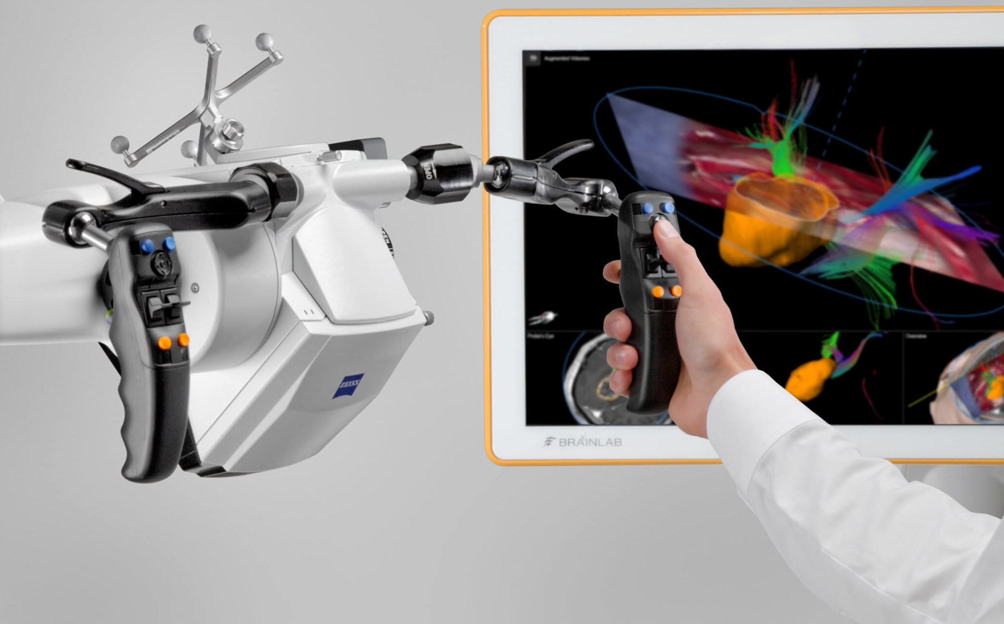
Brainlab invites you to join our live webinar, “Augmented Reality-Assisted Surgery – The Benefits of Microscope Navigation in Neurosurgery” on February 10, 2021 at 4:00 PM CET. Presented by Margrét Jensdóttir, MD (Karolinska University Hospital, Stockholm, Sweden), Sabino Luzzi, MD, PhD (University of Pavia and Foundation IRCCS Policlinico San Matteo, Pavia, Italy) and Christian Raftopoulos, MD, PhD (Cliniques universitaires Saint-Luc, Brussels, Belgium), this webinar will also include a discussion and Q&A session. 
The webinar will cover:
· Why and how to use augmented reality-assisted techniques in neurosurgery?
· The benefits of microscope navigation in tumor surgery
· The advantages of microscope navigation in vascular surgery
We look forward to meeting you online!
Participation is free of charge.
The views, information and opinions expressed during this webinar are the speakers’ own and do not necessarily represent those of Brainlab.

Palestrante

Margrét Jensdóttir, MD

Karolinska University Hospital, Stockholm, Sweden

LinkedIn Profile

Christian Raftopoulos, MD, PhD

Cliniques universitaires Saint-Luc, Brussels, Belgium

LinkedIn Profile

Sabino Luzzi, MD, PhD

University of Pavia and Foundation IRCCS Policlinico San Matteo, Pavia, Italy

LinkedIn Profile

To watch the webinar, please enter your contact data Please confirm that you would like to register for this webinar

    We offer our webinars free of charge. In return we ask for your permission to receiving promotional emails.

    Informações sobre o processamento de seus dados pessoais podem ser encontradas na política de privacidade da Brainlab

    Are you interested in an easier and faster way to register for upcoming events?
    Create a Brainlab ID account and save time before the next session. Gain access to MyBrainlab Online Services for more related event and webinar recommendations.

    Agradecemos por sua inscrição! Você pode visualizar o vídeo agora.

      To join our webinar free of charge, please confirm that we can contact you with related promotional material in the future.

      Information on the processing of your personal data can be found in the Brainlab privacy policy

      Thanks for your registration! You can now view the recording.

      Video Transcript

      Nadja Heindl: Well, good afternoon, everyone. Good evening, good morning, depending on where you calling in from, and very welcome to today’s webinar on “Augmented Reality-Assisted Surgery – The Benefits of Microscope Navigation in Neurosurgery.” First of all, thank you for taking the time to join us today. So, my name is Nadja Heindl, and I’m clinical marketing manager for neurosurgery at Brainlab, and it’s my pleasure to introduce our speakers for today and experts of the session.

      So, we have Dr. Margrét Jensdóttir here from Karolinska University Hospital in Stockholm, Sweden. Dr. Yenstir…Jensdóttir is also going to chair this session today. Her presentation will be followed by Dr. Sabino Luzzi from the University of Pavia & Foundation IRCCS Policlinico San Matteo in Pavia, Italy. And then last but not least, Professor Christian Roftopoulos from Cliniques Universitaires Saint-Luc, Brussels, Belgium.

      They going to share their experiences in using augmented reality in glioma and cavernoma surgery with you, and will also happy to answer your questions. Also with this in the webinar is my lovely colleague, [inaudible 00:01:17].

      Emese: Yes. Hello and good afternoon, and a very warm welcome, also, from my side. My name is [inaudible 00:01:24], I’m marketing manager at Brainlab. So today’s webinar is last around 45 to 60. Each of the three presentations will last around 10 minutes, and will be followed by a short Q&A session. If you have any questions, you can go ahead and enter them into the chat box on your webinar interface at any time during the webinar.

      The questions are going to be reviewed and can be discussed during the Q&A sessions. And in case you miss out on anything, don’t worry, the session’s going to be recorded and shared with you within the next week. And before I hand over control to Dr. Jensdóttir, let me introduce her more closely. She will then introduce the following speakers. So, Dr. Margrét Jensdóttir is senior consultant neurosurgeon, and head of glioma surgery and surgery and the awake surgery intraoperative mapping program, as well as head stereotactic laser ablations in her institution.

      Further, she’s a board member of the Swedish Neurosurgical Society, the National CNS Tumor Group, and the CNS Cancer Registry of the Swedish National Cancer Center. She has a special interest, both clinically and scientifically, in glioma surgery, and surgery…intraoperative [inaudible 00:02:41].and mapping, sorry. And with that, let’s get started. Margrét, the virtual stage is yours, and I’m going to hand over presentation to you.

      Dr. Jensdóttir: [inaudible 00:02:53] you very much [inaudible 00:02:54] presentation. It’s a [inaudible 00:02:57] to be had with my distinguished colleagues. And I guess you have all joined us here because you are interested in providing your patient with the best operation, and we are going to share with you our use of the navigated microscope in our practices, and we’ll see some different treatment cases for the low and high-grade gliomas, and finish off with challenging muscular case.

      And I assume you are seeing my slides, so I’ll just go on and continue. A short disclaimer, I am presenting my own experience and use, and a little background in numbers. I’m working at the…university hospital with the regional reference, and we are providing service primarily for the capital area and Gotland with a [inaudible 00:03:57] 2.5 million inhabitants. And in our department, the section of neuro-oncological surgery provides all modalities for neurosurgical treatment as well as the Gamma Knife and stereotactic laser ablation, and we do around 400 intracranial tumor resections a year.

      Well, we all know that the last decade has given you a mounting…given us a mounting evidence for the importance of extended resection, not…for the patient survival not only in high-grade gliomas, but also low-grade gliomas. And we have a lot of technical aids in [inaudible 00:04:38] to try to achieve maximal resection, like the navigation system, intraoperative ultrasound 5-ALA, and intraoperative MRI. But we also know that we can’t go beyond the functional port…border of the brain if we don’t want to inflict neurological deficits upon our patients.

      And we have also seen great evolution in technalities…technicalities to display these eloquent areas of the brain with fMRI, tractographies, and transcranial magnetic stimulation. So we are able to do meticulous preoperative planning to try to perform safe surgeries without damaging eloquent brain. However, we haven’t really seen, fact, all these different imaging modalities have been able to translate directly into the operating theater, at least not…or certainly not directly into the surgical [inaudible 00:05:47]. So, what we are going to discuss today is, for the last couple of years, improvement of the augmented reality.

      And with Brainlab’s microscope navigation interface, we are now able to implement and import all these planned structures, objects and tracts, into our ocular so we can have a…have it display not only [inaudible 00:06:14] visualizing all these structures on the navigation screen, but we can see directly into the microscope ocular. And I’d like to share with you one of the cases. This is a typical patient presenting with low…presumed low-grade glioma. A 29-year-old right-handed male who suffered from a first epileptic seizure.

      And we did show tumor, here is the FLAIR sequence, and we see that not only is there a small…there’s a small tumor part here in the insular, but it’s a larger tumor component postcentrally. And it caused some concern that on the T1 [inaudible 00:07:07] sequence, we could see small amount of diffused…diffuse enhancement in this area, and with perfusion, we also saw that there was more activity in this area. And since there seemed to be into tumor components being decided [inaudible 00:07:25] stage procedure, and go [inaudible 00:07:27] the larger tumor component, which was showing signs of some aggress [inaudible 00:07:36].

      So this is a screen from that navigation system, and that go into the OR, where you see a picture from our operating theater where we have that [inaudible 00:07:50]. The second screen is hidden here. And then we have the KINEVO microscope with reference store for the [inaudible 00:07:59], and we also using ultrasound for most of our glioma procedures. We have a Flex Focus, which also have calibrated probes for that Brainlab navigation system, [inaudible 00:08:10] you can see here.

      And then I wanted to show you, quickly, what it looked like intraoperatively, and in the first picture there, we were trying to see if this area with a little bit of diffuse enhancement, as you can see up here. This is the picture-in-picture feature of the microscope navigation, where you using the microscope as your pointer, and that pointing to this area, and we trying to locate the contrast enhancement with 5-ALA. We didn’t really get contrast enhancement in this…we didn’t really get any fluorescency in this area. But, as you could see in the beginning of the video, we were also doing a navigation, direct cortical stimulation, to make sure that we weren’t having any functional areas up here.

      And then we went further down to see if we could locate any fluorescence here to try to take biopsies from the most aggressive part of the tumor to get rightest pathological diagnosis. And if we speed up the video here a little bit, you can also see that we doing a navigated stimulation. You see the object overlay, and we having an…a electral strip there to see if we get [inaudible 00:09:45] charge while we doing the direct stimulation. And then down here, going for some of the tumor, and taking out the biopsy for [inaudible 00:09:57] there, from the fluorescing part.

      Moving on, we then go down with the…resecting the tumor anatomically, taking out the whole gyrus, and we didn’t detect any functionality. Then when we are coming, as you can see here in the picture-in-picture navigation, where the haircross on the video image is corresponding to the focus point, which you use as the pointer in the microscope. And pointing [inaudible 00:10:33], we be doing the stimulation [inaudible 00:10:35] dissecting the tumor all the way here, and doing the navigating stimulation in order to respect the functionality of the area. And then speeding it up to get out the tumor – ‘course you would wish you could do this sometimes in the OR.

      And then here, we get to the more interesting point – we are taking up the ultrasound to do an ultrasound [inaudible 00:11:05] stimulation. And here, I wanted to see you where you do a [inaudible 00:11:09] ultrasound, you get a 3D image. This is the preoperative ultrasound image, and this is after some of the resection. And we have [inaudible 00:11:19] it there to get back to the area where we could see a little bit of the tumor remnant here at the [inaudible 00:11:28] part. This is a sort of coronal ultrasound image and you see the [inaudible 00:11:34] spare tissue there.

      And with this, you could take the 3D image into the Curve Navigation integrated with the MRI, and more importantly, do a new 3D object of this ultrasound- enhanced hyperechogenic area showing tumor signal. And with that, update the roadmap. Here, we will jump right into that part where we were…had taken out the majority of the tumor and wanted to see if there was something left in the ultrasound. And now, as you can see here in the picture-in-picture, so this is all…this is the view I’m seeing [inaudible 00:12:23], then then I had the [inaudible 00:12:24] and I can continue the stimulation directly on the object and see if there’s any functionality.

      Oh, all right, we can continue to resect it, which we did with that suction with monopolar probe so we can do section and stimulation continuously – as you will see here later on. And this gives you the possibility to resect without having to look away from the microscope too often. Get your pointer pointing to the cavity, maybe have to move the microscope to see your images on the screen when you can get it all nicely displayed in the ocular. Finishing off with ultrasound to do a resection control, and this is what it looks like with the object there with [inaudible 00:13:30].

      Seems to correlate nicely with the area that is resected, and on the ultrasound you can see that the tumor remnant has now been replaced with reception cavity. And if you look at the post-operative MRI, the smaller tumor in the insular wasn’t the case for surgery now, but we manage to remove the variety of component. The post-[inaudible 00:13:58] component, they have no remnant approaching their small ischemic lesion in that more central part, but the patient did not suffer any neurological deficits from that. And then I would like to finish off to show you just a few slides from another tumor section to show some other features of the microscope navigation.

      And this is a pilocytic astrocytoma [inaudible 00:14:26] patient that was operated a year [inaudible 00:14:29] a year [inaudible 00:14:30] in a different center for a large intraventricular tumor, and there was a very small remnant in the [inaudible 00:14:39] part of the third ventricle. And since it was pilocytic astrocytoma [inaudible 00:14:45] W8…WHO grade 1, the patient did not get any adjuvant treatment.

      But with follow-up after only one year, we started to see them, the patient had move to our [inaudible 00:14:59] center and we started to see this growing lesion in the bottom of the third ventricle, so we decided to do an open biopsy and partial resection to get the [inaudible 00:15:11] pathology. And here, you can see how you can remote control the navigation of the microscope with the microscope handgrip, then you have this display, and you can choose which features you are using. For example, the picture-in-picture navigation, or if you want to have your object displayed.

      And to see here in the beginning that [inaudible 00:15:35] was good and we were…and you see where the haircross is in the picture, it correlates nicely with the surface. But when we started to get into the ventricle, you could see that something seem to have happened and there was some lateral shift. And go a little bit further and come down to the ventricle… And the reason I was using the navigation here was mainly because this patient has been operate before, I didn’t really know what to expect inside the ventricle. If it could a lot of adherences or fibrotic tissue interacting with [inaudible 00:16:36] of the tumor.

      So, when trying to get the focus on the [inaudible 00:16:42] part, you see that we are placed a little bit more laterally. And it’s maybe better displayed here on the screenshot from the navigation screen. So what you going to do then is to try to update the navigation because is the most challenging part for the navigation miss…of course, to maintain accuracy throughout the resection. And there is the feature where you can do [inaudible 00:17:08] the navigation with maximal [inaudible 00:17:12] of the [inaudible 00:17:14] where you can correlate your MRI image with the anatomical surface, but it’s limited since it’s difficult to compensate for a lot of vertical brain shift.

      But in this case, we could use the object instead, so we did a navigation update from the object. And with that, we [inaudible 00:17:39] better navigation. Now since we’re working in a really deep part of the ventricle and start to have the picture-in-picture navigation because it’s rather pixel where you come to the deeper structure, but you can see [inaudible 00:18:02] here doing that [inaudible 00:18:07] navigation update up from [inaudible 00:18:09], and that provided us. Again, we can see how we can use remote control to change between the different interfaces in the [inaudible 00:18:21] display, and then after the update, getting a better registration, a better roadmap.

      And then it was quite assuring to have, just as a reminder, that we were working on top of the [inaudible 00:18:40]. And [inaudible 00:18:44] it was possible to take out the majority of the tumor, it was [inaudible 00:18:49] ventricle. You of course had a tumor remnants in the more diffuse part. So, what are the benefits of the microscope navigation? Well, I find it gives you a very efficient and ergonomic workflow because you have the possibility to do a more focused resection without the interruption of taking in the pointer and moving out the microscope and so forth.

      And then you have the opportunity for augmented visualization of your object, of your tractographies. It gives you a better planning and spatial orientation, so I find it very useful both for the experience certain, but this gives you also opportunities to educate and show for your [inaudible 00:19:34] surgeons and trainees what the structures you are expecting to see next in the resection fields are, and so fort.

      So it’s really a good supplement to all your technical helps and aids you have in the operation theater to do the best surgery, with…give the patient the best surgery, the maximizing resection while not compromising their neurological function. And thank you very much. So, I will hand over that slide. Please, Sabino, go ahead and [inaudible 00:20:27] your slides. And while you are doing that, I will see if there are any questions that we should address right now.

      Dr. Luzzi: Thank you. Can you hear me?

      Dr. Jensdóttir: We hear you loud and clear. And there is one question here, is, “How much more time is needed for surgery?” Of course, like when you are implementing a new technique in your OR, you have to allow some time for training and getting used to the…to system. So, of course, I wouldn’t recommend that you start using it with your most challenging cases, but use it for your more straightforward cases, like metastasis and gliomas in the noneloquent areas, just to get used to the system and all the features.

      Because after that, you don’t need much more time for the surgery, it’s…it can even be so that you’re using less time, because in the end it will be more seamless and efficient. Well, I think will go straight on to your profess…your presentation, Dr. Sabino. And it’s a pleasure to [inaudible 00:21:42] introduce you. You are the assistant professor of neurosurgery in the University of Pavia, as well as a consultant neurosurgeon in the Hospital San Matteo, Pavia, Italy.

      And you have a special interest in skull-based surgery, vascular surgery, and neurological surgery, and are a faculty member of many well-known hands-on courses. And it will be a pleasure to hear your experience and the…see that how you are using the microscope integration in your OR.

      Dr. Luzzi: Okay, thank you. Can you hear me? Can you hear me, Margrét?

      Dr. Jensdóttir: Yes, I can hear you loud and clear.

      Dr. Luzzi: Thank you so much. Can you see my first slide, please?

      Dr. Jensdóttir: I see your first slide, it’s looking good.

      Dr. Luzzi: Okay. Thank you. Thank you so much, once again, to Margrét. And thank you also to Brainlab for this kind invitation about this special topic. And I also wanna thank all the participant for having joined us during this afternoon for this special topics. And I want to share with you now with my [inaudible 00:22:59] experience about the use of “DTI-Fluorescein Augmented Reality-Assisted Surgery in Dealing with Supratentorial High-Grade Gliomas.”

      First of all, I wanna stress the concept according to which from an extenstive analysis of the [inaudible 00:23:14] it [inaudible 00:23:15] that glioma surgery mainly have two important landmarks. The first one, the first concept is to try to maximize as much as we can our [inaudible 00:23:26] of these tumors, and that the second aspect is to try to do of our best to keep high the quality of life of this patient. In doing so, the surgeon has to deal with two important aspects, and namely an accurate planning and the perfect execution of surgery also, but above this aspect are base it on a very deep knowledge of the anatomy.

      When I talk about “deep anatomy knowledge,” I obviously mean brain anatomy, but also neurovascular anatomy. The planning is in one of the most important aspects for the sucks fest of this surgery. And always, always, we should keep in mind that decision are more important than incision. And I want to stress the concept according to which a planning is, once again, one of the most important part of this surgery. When I plan my brain tumor surgery, I routinely use different modules and function of my navigation platform, especially fiber tracking.

      Fiber trackling completely change my life when I started to use it, and always the analysis of fiber traiting…tracking algorithm and tractography during my planning of brain tumor surgery change my point of view about my approach and allows me to tailor the approach with specific type of lesion and the specific type of patient, also. Here, I have briefly summarize our acquisition protocol for augmented-reality surgery, and we interestingly use always our echo-planner, diffusion-weighted imaging a lot with deterministic dual-tensor trackie algorith for [inaudible 00:25:07] extraction.

      And here, I have summarize the…our early experience with this technique in dealing with high-grade glioma. Only supratentorial high-grade gliomas in [inaudible 00:25:19]. We started to use this technique in January 2019. We have treated 63 patients, and regardless from the location and those occurrence of the pathology, we achieved a [inaudible 00:25:32] rate of 84% percent in terms of exsensor…extent of a section of the lesion. And we also have average postoperative NANO score of 10.1. But let me show you some cases of mine.

      The first case is a huge cystic tumor is a postcentral gyrus glioblastoma ‘fecting the 43 years old female, left…suffering from a left-sided arm and face numbness. This is a nice case for me just to show you how the preoperative analysis of DTI can affect your surgery and can improve your surgery. Also, when I saw this images for the first time, I suppose that this tumor came from the motor area, primary motor area. So, I suppose that probably the best way to approach this lesion was anterioric, but after the patient and they went to preoperative DTI, I realize – as we see now, within few second – that the corticalspinal tract was completely push it forward.

      So it means that this tumor probably may a region from the postcentral gyrus said this is the typical pattern of growth of postcentral gyrus tumors, so I decided to approach this tumors from…this tumor from posteriorly. Here, we have also the opportunity to tailor our craniotomy, and in dealing with this region, we have to keep in mind that we always have two important landmark, especially two venous landmark. The first venous, the first vein, the first one is the anterior one, and this vein runs always within the central sulcus, whereas the second vein, the second one runs at the level of the postcentral sulcus, so it means that I had to work in between these two veins to take out my tumor.

      This is the [inaudible 00:27:28] injection into the ocular eyepiece of the microscope of the DTI imaging in the form of the augmented reality. Regardless, by the fact that I had to perform, as in this case, a [inaudible 00:27:43] approach, obviously this tumor reached a cortical surface, so it is a cortical-subcortical tumor. I always like to open the sulcus by mean of the sharp dissection just to identify my boundaries of the tumor before to touch the brain and before to proceed the tumor, and before to take out the tumor.

      Obviously, when we deal with this region we have to do any effort to spare cortical vein here because they may be associated with catastrophic [inaudible 00:28:11] here, and I’m just behind the omega region, just behind the motor strip. Whenever possible, that I’ll also like to use ICG videoangiography just to check the flow, especially in the venous compartment, but artery also. And this is the postoperative surgical field in exoscopic view, also. And here, the cortical representation of the corticalspinal tract, and obviously it varies according to the focus.

      And this is the postoperative MRI showing gross total resection of the tumor, along with perfect visualization of the corticalspinal tract. The patient was discharge on the third postoperative day neurological [inaudible 00:28:56]. This is the patient. But let me show you my second case. My second case is a completely different story. These are primary motor cortex glioblastoma involving the left dominant hemisphere in 65 years old female suffering from new onset seizure.

      As we see here, this is a completely different tumor because this is a smaller but more complex tumor, in my opinion. This is tumor lies exactly in front of the omega region, in front of the motor hand knob. So, it means that I have to change my approach for this tumor. I have to approach these sue…tumor by means of a [inaudible 00:29:33] approach. I can approach these tumors from anteriorly rather than from posteriorly, and according to the growth pattern of these tumor, it…they [inaudible 00:29:43] on [inaudible 00:29:44] stage.

      They tend to split the corticalspinal tract into two halves. The first half is push it anteriorly, whereas the second half is push it posteriorly. And also, the inferior [inaudible 00:29:56] of these tumor had intimate relationship with the arcuate fasciculus at the inferior part. And also, the dorsolateral aspect of the SLF1 or the…of the superior longitudinal fasciculus. Now I’m opening the precentral sulcus here, so everything behind is a motor area, whereas what is above and anteriorly is the posterior part of the middle central gyrus.

      Always I prefer to perform my sharp dissection here just to expose the boundaries of the tumor. The main reason why this patient wasn’t operated on [inaudible 00:30:40] surgery was the fact that at that time in my hospital I didn’t have the opportunity of fully trained team to perform [inaudible 00:30:50] surgery. Now I’m completed my dissection. Obviously, in this case I use cortical-subcortical mapping just to check the function, because one of the most important aspect, as I will say afterward, is the fact that we have to control function because DTI provide only structural anatomical information, but we have no information about the function of this lesion. And now I’m working on the tumor, obviously direct approach, piece by piece removal.

      Special care should be take to avoid any aggressive coagulation with this…within this area. And the last part of tumor was done under a constant check of the corticalspinal tract, and also are wake…arcuate fascigal, sorry. This is the postoperative field at the end of surgery. This technique can be combined also with ICG, as we see. And here, once again, some picture of surgery. Postoperatively, this patient suffered from right hand numbness, but she completely recovered after a third week…after three weeks, and third weeks follow-up.

      And this is the postoperative MRI showing, once again, a complete dissection of the tumor, along with a very good presentation of the corticalspinal tract, and also the arcuate fasciculus. Let me show you my last case. My last case is an insular tumor, a huge tumor here in 63 years old man suffering from hypermotor epilepsy. When we deal with insular tumor, we have to take into account as insular tumor is always a challenge in neurosurgery, is because insular tumor stand to push, medially, the corticalspinal tract and to intimate relationship with the optic tradition, the eye [inaudible 00:32:44], and also especially with this huge tumor with a [inaudible 00:32:47].

      This is the pattern of growth of this tumor that reach in at the level of the extreme external capsule, and whenever possible, I wanna stress that I prefer to use a pterional-transsylvian approach for these tumors for certain reason. The first reason, rise in the fact that pterional approach allows me to spare the opercular part of the frontal lobe, temporal lobe, and also parietal lobe. The second reason is the fact that I had two opportunity to avoid the rigid [inaudible 00:33:17], as we see in the video within few minutes.

      The third reason is that I have multiple walking corridor. And the fourth reason is the fact that I have a much more control of the [inaudible 00:33:34] here. I wanna stress that [inaudible 00:33:37] has nothing to do with insular tumors here, [inaudible 00:33:41] provide only for the vascular supply of the basal ganglia and [inaudible 00:33:45] internal capsule. The vascular [inaudible 00:33:48], the vascular [inaudible 00:33:49] the cortical surface came from the M2, M3 segment of the MCA, and the key of this surgery is a wide splitting of the Sylvian fissure.

      I have already opened the so-called “sphenoidal part” of the Sylvian fissure, now I just complete the opening and the splitting of the so-called “opercula insular part” of the Sylvian fissure, and I have just expose the [inaudible 00:34:13], the superior, inferior, and anterior limiting sulcus of the insular, along with the long axis of the MCA. Now I starting to remove the tumor. Please note that I don’t use a spatula here because I have already open in the fissure and I avoid any [inaudible 00:34:29]. Cavitron and with Transonic aspirator is very good for these procedures, and now this is the most delicate part of surgery and very close to the [inaudible 00:34:38] here.

      And I prefer to have a [inaudible 00:34:41] check of corticalspinal tract, and also the cortical-subcortical mapping to get more function. As already said, I like to check the flow at the end of surgery. Now we see the anterior temporal [inaudible 00:34:54] and those of the [inaudible 00:34:55], and this is the surgical [inaudible 00:34:57] at the end of surgery, also. This is the possibility of MRI showing a complete resection of the tumor, along with nice visualization of the [inaudible 00:35:06] of the corticalspinal tract. In conclusion, these techniques make the surgeon have…this technique have several advantages, in my opinion.

      But especially this techniques make the surgeon out…much more aware about the [inaudible 00:35:23] location of the [inaudible 00:35:24]. It should be stressedalso that the techniques had some technical constraints. The first one is the fact that DTI only provide for structural and anatomical, but no functional information, so that’s the main reason why it should be coupled with functional mapping techniques. And also, we have two further shortcomings. The first one is the parallax, and the second one is the crowding of the fiber tracts.

      They can be reduced or decreased by means of the [inaudible 00:35:52] going to [inaudible 00:35:53] function that significantly reduces the parallax, but also a strict [inaudible 00:36:00] selection of the white matter fiber tracts to deal with during surgery. That practically speaking, are no more than two or three for the so-called “deep-seated in high eloquent region.” In conclusion, DTI-fluorescein augmented-reality surgery techniques is safe and effective in maximizing the extent of resection of supratentorial high-grade gliomas.

      It allows for detailed intraoperative virtual topographic map of the white matter fiber tracts, and also, it allows for a safer surgery in the so-called “highly eloquent” areas. But let me stress the last concept according to which, despite the high occurrency of the techniques, has always set for me a perfect knowledge, a deep knowledge of the anatomies. A starting point to perform in any case, a [inaudible 00:36:49], and especially a safe resection of these tumors. Thank you.

      Dr. Jensdóttir: Thank you very much, Sabino, [inaudible 00:37:00] elegant and [inaudible 00:37:03] presentation. I think Professor per…Roftopoulos, if you could start getting ready with your slides, and we’ll see if we have any pressing questions where we can take them in the Q&A after your presentation.

      Prof. Roftopoulos: Yeah. [inaudible 00:37:30]

      Dr. Jensdóttir: Yes? Well, I…

      Prof. Roftopoulos: Can you see my screen?

      Dr. Jensdóttir: …[inaudible 00:37:33] Christian Roftopoulos, who has been the head of the… chairman of the neurosurgical department at the Saint Luc University Hospital in Brussels since 1996. And professor since 2000 at the same university, and he has, under his leadership, made the department grow internationally to a recommended center of excellence that attracts visiting colleagues and patients alike. And it would be interesting to see your challenging case. Please.

      Prof. Roftopoulos: Okay. Thank you to everyone. Thank you to Brainlab to have invited me for the presentation, it was a pleasure to be here. You see that the subject of my presentation is related to brainstem cavernomas, and I will stress the augmented reality of…that we use to reach our goal. First, I must [inaudible 00:38:48] the augmented-reality navigation of neuronavigation is a so clear a concept that I started to use it since 1996. And as you can see here, my first microscope allowing me to perform what I called “frameless stereotactic surgery” was the MKM system. It was a wonderful piece of machinery, and sometime I miss it.

      But, even if we are performing augmented-reality surgery since 1996, the augmented reality has gone through everything in our life. And for example, I’m using also augmented reality in my car since five years, and you see that what computer can give you as information to add, to superpose through the [inaudible 00:39:51] is a wonderful phenomenon, development in the [inaudible 00:39:57]. So, most operation – and I would say all operations require perfect preoperative preparation – and you see here what I call “preoperative virtual surgery.”

      We started to develop that context more than 13 years ago using the Destroscope. You see the Destroscope with Google…with goggle, show you the brain of your patient in the space in a 3D virtual volume, and you can perform on that head, virtual surgery. Other people have developed that concept for aneurysm surgery. You see here a surgeon training himself with also goggle to have 3D view of the augmented reality of his patient, and that can give to that result where you see your clip and you see the formation of the virtual aneurysm when you are performing that virtual clipping.

      You see here one of my colleague performing a preparation on the Destroscope in the year 2007, more than 13 years ago. I would like to illustrate my comment using a case. It’s a case of brainstem cavernoma, it’s a [foreign language 00:41:44] cavernoma. And I’ve decided, looking at the MRI, to perform heterolateral approach. You see it’s a huge one. This cavernoma has bled many times, and at the end, the patient decided for surgery even if we stress the risk related to that kind of surgery. We decided for contralateral approach, and as you can see, we have identified different volume of interest.

      You see the target there, actual section showing that you…inferior part of the cavernoma is going to the [foreign language 00:42:37]. You see here, clearly, the omega, the target in green, and you have the phonics in yellow, and in blue you have the internal cerebral vein. For me, it was enough to proceed to surgery and to perform a mini mild invasive surgery. It’s one of the advantage of using augmented-reality surgery is to be more minimal invasive, even if you are performing a very deep and potentially aggressive surgery. You see, for the surgery, here we are in the operative position. Is on the right side.

      And here, I have my virtual trajectory. And I have to stress different point, is first, SL is the “septum lucidum.” I have already think when your surgeon [inaudible 00:43:59] their work, their way in the cavum, into the septum lucidum. And you know that in this case, some anatomies describe a cavum of the septum lucidum interior part, or cavum vergae in the posterior part. Anyway, it’s a cyst inside the [inaudible 00:44:26] of the septum lucidum, and you have to be aware of that. The second thing is the phonics. Really important. The phonics, which is hidden by plexus choroid, and a little bit below you have the internal cerebral vein.

      And you know that to reach the third ventricle, what you will have to do is to go through [inaudible 00:44:55] on the phonics to push to the midline velum interpositum, to put the midline the [inaudible 00:45:05] vein. And so, you will reach a third ventricle and [inaudible 00:45:14]. Here, the [inaudible 00:45:17], the augmented-reality navigation, has help me tremendously to identify, especially the point of entrance into the [inaudible 00:45:32] of [inaudible 00:45:33]. So you see on the left side, the surgical position. In green you see the target, the tumor that I will have. The tumor, yes, it’s a kind of tumor, vascular tumor.

      You see in below the [inaudible 00:45:53] vein. No, I’m opening the septum lucidum to reduce the pressure inside it. And so I have a better [inaudible 00:46:03] visualization of the social [inaudible 00:46:07] the thalamus, I see the plexus choroid. I know I’m decide…bisecting the choroidal fissure, and you will see that I’m go…I’m into the third ventricle. And I [inaudible 00:46:23], trying to [inaudible 00:46:24] on the medial surface of the thalamus something a little bit orange. You see some…somes…the intra-[inaudible 00:46:35] vein on the left side.

      No retractor, only pushing, running back, pushing, coming back. I’ve gone through the medial phase of the left thalamus, and I’m starting the dissection of this huge brainstem cavernoma. You see my opening is minimal. On the right side, the thalamus, the right thalamus, left internal cerebral vein, the velum interpositum on the left side, and slowly but surely you have to extract this huge brainstem cavernoma and try not to perform any electrocoagulation to avoid any ischemic problem. You see it’s not so easy to push, to extract the lesion through the operative pathway. I will divide the brainstem in small…in smaller pieces to be able to remove it.

      The last one is here. I know I will check my hemostasis, and to be sure that no remnant of the cavernoma stays into it. What is really important is not to remove the gliosis, because gliosis is not anymore brainstem cavernoma. So, I inspect my operative field, we are [inaudible 00:48:30]. I remove [inaudible 00:48:31] from the [inaudible 00:48:32] to avoid any contamination, excessive contamination, of my ventricle system. You see the opening, less than 20 millimeter in front of the central sulcus. The patient the night of the [inaudible 00:48:48]. And you see I was impressed that he was so well a fewer hours after surgery.

      So we are discussing with him in English [inaudible 00:49:14]. And here, you see five days after surgery. Okay, of course I have to show you the postoperative image. You’ll see a complete resection. So my messages, what I would like that you remember regarding this presentation is, that regarding brainstem cavernoma, the first thing is, of course, the strategy. You have to think about the direct, or even if a contralateral approach, please try to avoid the fourth ventricle floor because is very delicate. The second point, don’t hesitate to use augmented-reality navigation.

      In other cases, which has been published in [inaudible 00:50:20] neurosurgery, the use of tension, of diffusion, of…to identify the different tract are also very important. I do not choose intraoperative monitoring. Gross total resection, fortunately, was possible in all the case in our series, and over 19 Casey…cases. One of the most important point is the frontier between the cavernoma and the gliosis. There, navigation won’t help you. It’s your experience you have and the quality of your microscope to identify this frontier.

      And the results, the patient were stable or better in 84%. Unfortunately, one of them develop rapidly a severe Holmes tremor, so that this series do not show 100% stable of better clinical status. Holmes tremor, also named “rubral tremor,” is very, very special because it doesn’t develop immediately after surgery. It developed one, two, or three weeks after surgery. That means that the patient is a little bit degraded after surgery but recover, and you think that everything is going well, and a few days later, the patient developed this tremor. One of the patient was develop such a tremor was treated very effectively by brainstem stimulation. I would like to thank you for your attention.

      Dr. Jensdóttir: Thank you very much, Professor, for an elegant illustration of a very challenging case. And for you in the audience, feel free to send your comments and questions, and we’ll try to address them all the best we can. But I could start of by asking you, Sabino, when you are planning your high-grade glioma cases, and that especially in the OR, are you using the integrated navigation all the time? That the objects displaying in [inaudible 00:53:06] time? Or do you have any comments on that? Because there a lot of people are asking if this is just like blinding lights if you having it interrupting your visual field?

      Dr. Luzzi: Can I reply to you [inaudible 00:53:25]?

      Dr. Jensdóttir: Yes, please.

      Dr. Luzzi: Okay, yeah. I spent a very long time for my planning, sometimes even days, because I try to realize, for me, anatomy’s everything. So, I need to have as much [inaudible 00:53:42] information as I can by a singular case. I review my diffusion-weighted imaging and I review my FLAIR imaging because I want to realize in my mind, precisely, [inaudible 00:53:55] of the tumor. And then I go into the operative room and I reconstruct, I try to perform my fiber tracking, and I then navigate my fiber tracking. Navigate, navigate, navigate. And then I realize my surgical route and my surgical corridor.

      For me, I have a concept that…but is not what…my idea, it is a universally [inaudible 00:54:17] idea, according to which for me, minimal invasive surgery doesn’t mean a small skinny incision or a small craniotomy. Sometimes I make very huge craniotomy but the patient stay very well. For me, minimal invasive surgery means a perfect anatomical respect of the neurovascular structure of the brain, of the lobes. And I like to stop exactly what I had to stop surgery at…for a deep-seated lesion I always to prefer [inaudible 00:54:48] approach because I realize my trajectory to that kind of leisure and the specific kind the patient.

      And for more, as already said, fiber tracking completely changed my life because I have the opportunity to realize… Obviously, I have to stress that their concept according to which DTI is not [inaudible 00:55:10] is not the real life because a… So, you know, we have some question about the fact that [inaudible 00:55:18], the angular selection of the probabilistic reconstruction of DTI in technique [inaudible 00:55:24] also, algorith [inaudible 00:55:27] anyway. It help me, and it help in my daily practice for choose my best approach.

      Dr. Jensdóttir: Thank you very much for your answers [inaudible 00:55:39]. And I totally agree with you, minimally invasive can mean more than just a small incision. It’s very important to be sure about the [inaudible 00:55:49]. And like you show this, also, elegancy, Professor Roftopoulos, in that your approach, with no traction and especially no vascular damage in the lesion. But I have a question that, when you are controlling the microscope, because a lot people ask that, how you control the different functions in the microscope? Are you using the handgrips, or do you use, like, the mouthpiece or [inaudible 00:56:21]?

      Prof. Roftopoulos: No, my foot, my right foot is very… Yeah, you have everything with [inaudible 00:56:29] surgery. We do not speak enough about [inaudible 00:56:33] surgery. There’s a reason why you can perform a very deep surgery with a small opening. We know that craniotomy takes time. Huge craniotomy increase the risk, that there has been very well published. With augmented reality, you can recognize the different [inaudible 00:56:57], so…and if you want to go fast, I really think that one of the fastest way to manipulate your microscope is with your feet.

      Dr. Jensdóttir: And that comes directly to a question from the audience here, where they’re asking about the…a general question about robot assisting…assisted neurosurgery, and we haven’t really addressed that matter. There are some elegant functions where you can use the robotics to…the integration between the microscope and the navigation to get the microscope into a position where you are defining a point in your surgical field, then you can let microscope memorize that trajectory, for example.

      But also, that this gives you the opportunity not to concentrate on your navigation screen. So you’re not really having to concentrate on the [inaudible 00:57:59] because you can make the whole procedure through your microscope, and getting all this information integrating in your visual field. And like Professor Roftopoulos said, controlling the microscope with your foot, that’s give you a lot of advances, then your…you never have to [inaudible 00:58:20] instruments in your surgical field.

      Professor Roftopoulos: But [inaudible 00:58:25] robotic, we have to define the robotic surgery. The MKM, more than 20 years ago, was robotic microscope. And so, you push on a button and the microscope put itself in the batter…in the pathway that you have define. But it’s not really useful because with Penteho [SP], with the last version of the Zeiss microscope, coupled with Medtronic, of Brainlab, has…in our department navigation you find the pathway so easily. I think that robotic microscope is not economic efficient for the future, and I think it’s the reason why Zeiss do not invest in that kind of development.

      Dr. Jensdóttir: Yes, thank you. And I find it most [inaudible 00:59:25] in a game-changing community just to be able to use the navigation, the picture-in-picture navigation, where you can navigate with a microscope so you don’t have to take the pointer and see if you getting close to the [inaudible 00:59:40]. Especially when you are operating…removing low-grade gliomas, where you are practically not seeing as that many…much differences in the tumor tissue in the surrounding brain, so you get…constantly have the assurance with your navigation in the ocular that you are moving in the right direction.

      And that can also be useful in the high-grade gliomas, because as you all know, it’s not the tumor core itself that’s difficult, but it’s the core, like you mention Sabino, that always keeping track of where you have your tractography. And that when you moving out of the necrotizing, more solid part of the tumor and into diffuse border of a high-grade, I assume you use the same technique, that always keeping track of where you are, especially concerning the tractographies. Or do you have any special comments on that, Sabino?

      Dr. Luzzi: Yeah. I completely agree with you. And I am strongly convinced that in the near future, probably the key will be in the ultrasound because I really appreciate your cases operated with ultrasound. Because we have a [inaudible 01:00:59] information, because it is not only an updated information, I routinely use it to operative room, a KINEVO microscope. I also use Curve Brainlab but I routinely use my correction of the brain shift self-alignment, but I have to keep in mind that that is not the real-time navigation.

      The only form of real-time navigation you see ultrasound, so I very like ultrasound. That my congratulation for your case, yes, that are very nice, and also the case of Professor Roftopoulos, so my best. And for me, I completely agree with you.

      Dr. Jensdóttir: There is one question here regarding the difficulty level for removing low-grade glioma. That’s, in fact, a challenging question to answer because, well, as we have stressed upon, the difficulty is in appreciating where you have the functional border and where you have the eloquent areas. And that is also as challenging in other regions, but with low-grade glioma you have this added challenge of the tissue not being so much different from the healthy white matter. But as Professor Roftopoulos mentioned, it’s…it takes a lot of experience, of course, to really appreciate all these eloquent areas in the brain that you have to preserve, and to use all this information in the best way.

      But that’s why I would say difficulty level is, of course, for more experienced surgeons, like with all these cases we have presented here today. But it doesn’t mean that you couldn’t be using the navigated microscope and all these advances of the augmented reality because…and like every tool, you need experience, you need training, so you should use it with all your routine cases – with gliomas, in noneloquent areas, and so forth. And we are getting a lot of comments that people really appreciate it, and I want to thank you, Professor Roftopoulos and Sabino Luzzi, for your elegant presentations.

      And I don’t know if we have more time for questions, or if we should also thank Nadja and [inaudible 01:03:49] for planning and making this webinar possible. And I know there’s been a recording for those who couldn’t join us here today, for…and prob…maybe answer some more questions or comments if they are.

      Emese: Yeah, we’re a little bit over time, so I think with that, we can finish up this 65 minutes of wonderful presentations. We want to say thank you to our three wonderful presenters, we very much appreciate the time you invested and all the insights you shared with us today. And also, thank you to the attendees for the interesting questions. Those questions which we were not able to cover now within this session now, we will follow up on via personal email.

      And, yeah, as Margrét mention, the recording will also be sent out to you within the next couple of days. Okay, and I think with that we can finish off. And for now, we wish you a great rest of your day. Stay safe, and goodbye. Thank you, everyone.

      [crosstalk 01:04:57]

      Emese: Thank you, Dr. Jensdóttir, Professor Roftopoulos, and to Dr. Luzzi, thank you.

      Dr. Luzzi: And a special thanks also to [inaudible 01:05:03] and Nadja for the special technical support. Thank you so much once again. All my best.

      Emese: Thank you all. Thank you, same to you.

      Dr. Jensdóttir: Thank you.

      Emese: Bye.

      Prof. Roftopoulos: Bye-bye.

      Dr. Luzzi: Bye.

      Emese: Goodbye.

      Dr. Luzzi: Bye.

      Related Webinars

      02Blurred_lights
      Augmented RealityNeurocirurgia
      SEEG Planning Integrating Automatic Anatomical Segmentation and Augmented Reality

      Brainlab invites you to join our live webinar, SEEG Planning Integrating Automatic Anatomical Segmentation and Augmented Reality, on September 14, 2020 at 2:00 PM (CET). Presented by Dr. Peter Reinacher, Neurosurgical Consultant of the Department of Stereotactic and Functional Neurosurgery …

      Dr. Peter Reinacher
      186001594307693
      Augmented RealityCraniomaxilofacial+ 1
      The Role of Mixed Reality in Craniomaxillofacial Surgery – An Interactive Discussion about Our First Experiences and the Future Outlook

      Brainlab invites you to join our webinar, “The Role of Mixed Reality in Craniomaxillofacial Surgery – An Interactive Discussion about Our First Experiences and the Future Outlook”, presented by our four international speakers: Bradley Strong, MD, Professor and Vice Chair …

      Bradley Strong, MD
      Bernd Lethaus, MD, DDS
      Alexander Bartella, MD, DMD
      Reinald Kühle, MD, DMD

      See more upcoming webinars

      Register now