Webinar

Benefits of navigation and intraoperative imaging in Cranio-Maxillofacial Surgery – a clinical case review

Topics
術中撮影
CMF
外科手術
手術ナビゲーション
言語

English

Share this page

概要

Brainlab invites you to join our live webinar, “Benefits of navigation and intraoperative imaging in Cranio-Maxillofacial Surgery – a clinical case review”, on February 9, 2021 at 4:00 PM CET presented by Prof. Julio Acero, MD, DMD, PhD, President of the European Association for Cranio-Maxillo-Facial Surgery, Head of the Department of Oral and Maxillofacial Surgery of the Ramón y Cajal University and the Puerta de Hierro University Hospitals in Madrid, Spain, and Professor of Surgery at the Alcala University of Madrid.


In this webinar you will:

  • Understand the benefits of virtual planning combined with navigation assisted surgery in craniomaxillofacial surgery
  • Be familiarized with the workflow concerning the use of navigation and intraoperative imaging
  • Learn how intraoperative imaging can help to improve outcomes in complex craniomaxillofacial procedures

We look forward to meeting you online!

Language | English

In case you can not join the webinar, it will be recorded and shared afterward.

Participation is free of charge.

The views, information and opinions expressed within this presentation are from the speakers and do not necessarily represent those of Brainlab.

Speaker:

Prof. Julio Acero
Prof. Julio Acero

MD, DMD, PhD, President of the European Association for Cranio-Maxillo-Facial Surgery, Head of the Department of Oral and Maxillofacial Surgery of the Ramón y Cajal University and the Puerta de Hierro University Hospitals in Madrid, Spain, and Professor of Surgery at the Alcala University of Madrid

Moderator:

Jana Neider
Jana Neider

Product Manager CMF / ENT, Brainlab

Video Transcript

Jana: Welcome to our first cranio-maxillofacial webinar in 2021. My name is Jana Neider, and it’s a pleasure for us to have you online here today. Before I introduce you to our speaker, I would like to explain a few points. Today’s webinar focuses on the benefits of navigation and intraoperative imaging in CMF surgery. It’s a very interesting topic and has been asked many times in the past webinars, so I’m very happy that we have this as a topic today.

The lecture will last about 45 minutes, followed by a 15-minute question and answer session. Questions can be submitted only through the online chat function and will be collected and selected by me to be addressed to the speaker at the end of his presentation. The webinar is live, and it will be recorded to be watched again by all registered participants. For further questions, feel free to also use the chat function.

Now, onto our speaker, Professor Julio Acero. Thank you for being here with us today.

Prof. Acero: Thank you.

Jana: Professor Acero is most likely known to every one of you as president of the European Association but also from a lot of other associations. He is the head of Department of the Oral and Maxillofacial Surgery of the Ramón y Cajal University and the Puerta de Hierro University Hospital in Madrid, Spain. Furthermore, he’s known very much as a full professor at the Alcala University Hospital, and especially known to Brainlab for his tremendous knowledge in oncologic ad reconstructive surgery. Furthermore, he is using a lot of different technologies for pre-operative planning, navigation, as well as intraoperative imaging. His dedication to education will be very beneficial today because I’m sure we will learn a lot of new things from him today in this webinar.

We look very much for your presentation, Julio, and now the virtual stage is yours.

Prof. Acero: Thank you, Jana. It’s really a pleasure to be here sharing this time with you. And I would like to thank Brainlab for giving this opportunity. I think that the cooperation between the surgeons and the industry, especially an industry like Brainlab who specialize in high technology and offering us surgeons so nice tools for [inaudible 00:02:32] patients is really excellent.

So, my apologies also for having postponed for two or three weeks this presentation because as probably…I don’t know if you know, but I would say that I was affected by this horrible pandemia. I was hospitalized, and we were able to manage that. And so to postpone the presentation [inaudible 00:02:57]. So I hope that we will able in this hour to share our experience concerning the benefits of navigation and also the intraoperative imaging because I think that this is a tool that we can really use for the benefit of our patients. So, one second. I don’t know why it’s not running now. I will restart. Okay.

Jana: That’s good.

Prof. Acero: So, the purpose of our presentation is purely scientific. I have no conflict of interest to declare. Sorry, because something that we have previously…this, but I don’t know what…

Jana: We can see and hear you.

Prof. Acero: Yeah, I don’t know why it’s not running like it was, but I hope…Okay. So, as Jana said, I am working in Madrid in Ramón y Cajal and Puerta de Hierro Hospital with the University of Alcala, which is one of oldest in Europe. And I’m leading a young team which specialize in the different areas of our specialty. And so, in this presentation, we will try you to be familiarized with the workflow concerning navigation and intraoperative imaging, but also to understand…I know that it was also a topic of other presentations of virtual planning combined with navigation assisted surgery and learn how these tools can help us to improve our outcomes considers all our patients. This concern, especially in the bone surgery, cranio-maxillofacial surgery.

So, we probably have to consider that cranio-maxillofacial trauma was the area where this type of amazing tools were started to be used, but to summarize, our goals of treatment clearly is to get a good aesthetic outcome for our patients in every field of this specialty, in oncologic surgery, in trauma, and also in maxillo-facial deformities. In this sense, our ideal [inaudible 00:05:24] will be to show one of our post-operative cases like this and not to be able to identify what type of operation we have done. This patient was submitted for a maxillectomy, was planned under virtual planning [inaudible 00:05:42]. But what I want to show you, our goal is to get, in every field of our specialty of cranio-maxillofacial surgery or head and neck surgery, in general, because this also concerns other specialists, to get this functional [inaudible 00:05:58] result and also the cure of the patient in case of oncologic cases, of course.

So, it is clear that we have to have a plan. We cannot go to the operating room without a plan like it was in the old time. it was said by Benjamin Franklin, if you fail to plan, you are planning to fail. And, sometimes, our results or the results of cases that we see for other departments you have to say, but also we have complications. One of my, for me, better presentations we want about complications in reconstructive maxillofacial surgery, we’ll have to [inaudible 00:06:38] that we all can have complications.

And to avoid complications, start before the operation in the planning. And to avoid this type of result, we can have an oncological [inaudible 00:06:50] surgery, or in trauma, or after orthognathic procedure. So these bad results or complication, you have to have a plan. Miguel de Cervantes, a Spanish writer had said, “The man who is prepared has his battle half fought.” But also, Sun Tzu in “The Art of War” mentioned that the general who go to the battle with calculations before the battle will win the battle. And not so many years ago, I am not so old that I have to say that our planning not so many years ago was based on the [inaudible 00:07:27] and no clinical exploration, and we didn’t understand in a three-dimensional way our patients. But now, we are fortunate that we are in the future. The future is coming every minute, but it’s so quickly the evolution that we are in the future.

We are in the digital era. The digital age is an age which is defined as how the information can be handled, stored, printed, transmitted, and manipulated in many different ways that can help us medical imaging, which is defined like the DICOM phase as you all know, to manipulate, to manage our data, to share them. And this has opened a new world, let’s say, the world in which we are planning. So we have to become digital. The young people, our young colleagues, their friends, you are in the…and I’m very happy always working with our trainees and even specialists as you all know, and you are a people who were born in digital era. But people who were born before digital era has to become digitized. This is clear.

And digital information started to allow us to perform the diagnosis and planning of our treatment where we’re able to perform a patient’s evaluation in tumor, trauma, deformity in a 3D way, and also to plan our treatment. So, a preoperative planning based on the digital information opened, as I said, a new world and allowed to perform the virtual planning for oncologic resection and reconstruction, the planning of surgical correction of maxillofacial injuries and deformities, and the virtual design of reconstruction like flap geometry, or the design of customized plates or patient-specific implants. But if you think about that, this is theoretical in 3D but this is bidimensional. And this is a pre-operative planning, but this is not enough.

Failure in planning can lead to complication of results, but once we have our planning, we have to execute the surgical procedure and we have to counter devolution and to counter the results. Eisenhower said, “Before a battle, planning is everything. Once the fighting has started, this is worthless.” So, a good planning without a correct execution and the correct…or post-operative could be immediately post-operative control is nothing, and we can have also complications.

And for integrating all of these steps of the surgical procedure, pre-operative planning, surgical execution, and control of the results, we have a variable, as I said, amazing tools. And our operating facilities look really more complex than this operating theater of the “Avatar” movie. This is our own dedicated operation room we are sharing with the neurosurgeons not only the most advanced technology in imaging but our navigation system and our integrated intraoperative CT.

So, from planning to surgical execution. This started transferring virtual planning into clinical reality through stereolithographic models, 3D printing, templates production, surgical guides. These are tools that kept us, once we have planned it virtually, to go to the 3D reality and to help us to transfer our plan into the reality. And this is working for middle [inaudible 00:11:26] reconstruction, for a [inaudible 00:11:27] reconstruction, craniofacial, or orthognathic surgery. We can transfer the virtual planning of our post-operative situation through this type of tools in implants, especially patients with implants or with virtual guides.

But, moreover, execution of surgical procedure can be accurately guide by navigation. This is navigation assisted surgery, and we will discuss and we’ll show you how navigation helps us also to improve the transferring of the virtual planning into the reality of the OR and control of surgical results. What we have done is nothing we cannot check if it’s what we really aim to obtain. Traditionally, clinical evaluation and post-operative imaging was the method that we could check that our objective was good.

But, currently, we are able to introduce the intraoperative imaging immediately in the operating theater to check the immediate post-operative situation, especially in bone surgery. Of course, managing the soft tissues or magnetic resonance and CT is also accurate, but especially for bone surgery, we are [inaudible 00:12:48] soft tissues for bone surgery, so we have now a tool that can give us the possibility not to wait for the patient out of the operating theater to get a CT and to say, “Oh, my God, this was not what we wanted to have,” but we can do intraoperative, and this a facility the possibility to correct our error results immediately.

So, let’s discuss both concepts. Navigation, you all are familiarized, and many previous [inaudible 00:13:18] were based on navigation. Navigation is to correlate the patient’s anatomy with a previously downloaded data of the anatomy. So, a navigation is referring the patient to anatomy. And so this will allow the surgeon to see the position of the patient in the operating field in relation to the patient’s imaging in real-time.

So what is our workflow? This is based on the digital files. Digital files, once acquired, this is image acquisition, has to be downloaded into the system. And once the patient is in the operating theater, it has to be related to this datas in real-time, also allowing the patient to change the positioning, keeping the references. And once everything has been established, we can navigate what we have.

And this started not so many years ago, and as I mentioned starting this lecture, we started with facial trauma. This is a paper, one of our Chinese colleagues see how dedications for navigation were widening and also going to tumors and also for TMJ surgery. So it means that navigation is a tool that can help us in many circumstances. Our OR facilities, as I mentioned, where the navigation system integrated so well that was used normally is iPlan Cranial [inaudible 00:15:01] for our neurosurgical colleagues. We have a scale-based unit, and we are working very closely. We have also the tools to refer our patient to the system through the infrared light camera, the reference array, the pointer to mark the patients’ references, and the integrated AIRO CT scan.

So, going on in the, let’s say, review of the workflow, first, as I mentioned, is image acquisition. And so we have the diagnosis and we have the knowledge of the plan for patient. This case, I selected a trauma case, which is, let’s say, one of the prototypes of the indications of navigation assisted surgery and intraoperative CT in our field, and this is a case of orbital roof fracture. So, diagnosis, we know how is the patient, what is important. Virtual planning, we do it in the work station, and, normally, what we do is through segmentation of the unaffected contralateral orbit, we can have a model which can be transferred to the affected side through mirroring and be adjusted. I know that this can be discussed because not all patients are symmetric.

It could be it has been published a reference of, let’s say, libraries of orbits or an anatomical structure depending on the mean of the population. But, anyway, we have to look for a standard reference, and, normally, in our department, we choose the contralateral side to, let’s say, define what will be the idea of reconstruction. So once we have, let’s say decide what will be the [inaudible 00:17:04] we decide what type of repair of the fracture will be. In this case, we selected 8 to 10 images on implant, which is taken of the system library. In other cases, we have to prepare a patient’s specific implant.

Anyway, we have now a plan, and now comes the second step which is to transfer this plan to the OR reality. And, as I mentioned, the third point of the workflow is to refer the patient into the system. And these different methods, one could be the surface mapping, I would normally choose different points in the face to define, let’s say, the anatomical key points to be related to the information in the computer. So once we have calibrated the…correlates in real-time the patient anatomy with the pre-operative CT, or MR, or both, which can be previously downloaded, we have registered the patient.

Now, we can start the operation. And during the operation, depending on the pathology and the objective that we have, navigation can help us to check what we are doing, where are we, and especially this is really a very useful tool in midfacial surgery with an example of midfacial trauma. But also, and especially in oncologic midfacial surgery of skull base is critical because of the complexity of the anatomy. So we can see in the system where we are with the pointer partly where we are. Where are we, let’s say, placing our implants or replacing our bones in areas that normally cannot be seen by our eyes? So, intraoperative navigation in the three fields that we are considering can help us to check if the correction of the fracture is correct and if the placing of a plate, of an implant are correct.

In case of oncologic surgery, we can check the designed resection margins, and we will see other details that can be really helpful in oncologic surgery. And up in a new field in orthognathic surgery, we are now checking the osteotomy lines and also the position of the jaws and the movement. In my presentation with a clinical case showing you what we are now studying concerning navigation and intraoperative CT also in orthognathic surgery in my department.

So, these are two examples. This is navigation of a correction of the [inaudible 00:19:43] and this is a oncologic case. As you see, if you are familiarized with the facial oncologic surgery, you have to perform in a section without seeing the deeper structure because, normally, the maxillary bone is covering the tumor. But now, guided on the navigation, we know every time where we are, and this is really amazing.

But you all have to be aware that these are wonderful tools very precise, very accurate, but the surgeon has always, at least at the present stage of knowledge, to keep, let’s say, the contour. Because, sometimes, and this was…I don’t know if it’s fake news or something what happened, but it was in the newspaper how a lady from Belgium was going through the train station and finally ended in Croatia because she was following the navigation, the GPS of the car. And this car may be not so [inaudible 00:20:51] mistake, but who hasn’t had this experience? Like, it happened to me once with my car to be always trying to cross [inaudible 00:21:02] when the GPS system said, the navigation system in a car said, “You have to go through,” and this was strictly forbidden. Anyway, this happens. So the surgeon has to keep the contour.

And what happens sometimes? We are working, sometimes the reference start can be not so fixed, or something can change, technical mistakes can happen. This happened to us last week. You can see we are theoretically touching the bone with the pointer and the pointer was outside of the skull. So we have here a difference of free-forming images, but this is critical. In the area we are working goes through the optic nerve the intracranial carotid artery. So we have to check that everything’s accurate and know the benefit of the intraoperative CT is not only to check intraoperatively what happens but also the intraoperative CT has the capability to correct this.

So, summarizing benefits of intraoperative CT, in our view, are to check the final result in every type of bone surgery, to check the resection margins in tumor surgery, and superimposing the preoperative CT and the virtual planning we know that we got what we wanted. And this can be done in real-time and we can correct because the patient is still in the operating theater under the general anesthesia and we can correct something with what’s wrong.

And this is our case that we have been following to show you our workflow. You can see here, let’s say, the red image is our project, what we have planned. And we have navigated to put here a mass, and you can see the superimposed. This was an old mass, which was in an incorrect position, and here, the superimposal of our STL file of our project with the intraoperative CT which [inaudible 00:23:10] is showing that the positioning of the implant has been perfect. This is an orthognathic case that we are checking that the position of the upper and the lower maxilla are correct and are really merging or superimposing 100% with the previous plan.

And what about resection margins? Now, we are considering general workflow with these examples and the benefits of intraoperative imaging as the last point of our, let’s say, workflow in these cases. You can see here, it’s just highly sophisticated but I will explain you. We had a relapsing tumor which was a hemangiopericytoma in the skull base between the orbit and the brain. It was a relapsing tumor. Where’s the tumor? This is small two nodes because the giant tumor was removed three years ago. This is a benign aggressive tumor.

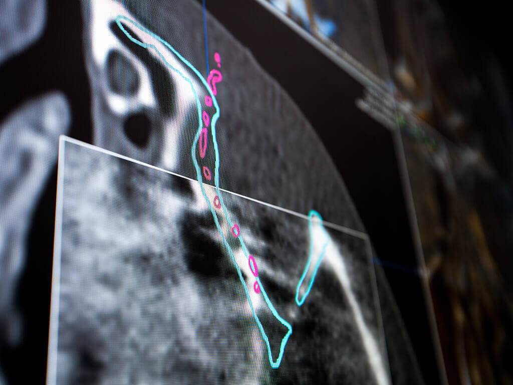
So what we are doing here, once we have resected in the navigation the tumor, we took an intraoperative CT, which is what we are seeing. Here you can see the tube and also the axial, coronal, and sagittal slices [inaudible 00:24:34] prints, and here is the tumor. It means that this was the previous imaging, but now in the intraoperative CT, there is nothing. And you can see that the bone margins are wide enough. So this is a method to check that we got adequate surgical margins in a complex relapsing tumor in this area.

And as I mentioned before, also the intraoperative CT, in case of losing the references that we mentioned before automatically can co-register what we have previously reused there in our system. If we lose the references intraoperatively and we cannot, again, perform the registration, taking an intraoperative CT, which is this case in another skull-based tumor, automatically, the system could register the situation with the previously registered information. It means that we can continue the operation and we have recovered, as I mentioned before, the accuracy of this situation.

So, what happened here? Let’s back here, and we are here. I hope this works. It worked. Yeah, this is the logistics. It’s not easy. You can imagine having an integrated system during the operation. Let’s say the anesthesiology has to place a respiration system, and everything has to be prepared also, the instruments table to allow the patients to be turned and to introduce the head in the CT. We have our dedicated radiologist. They come to the operating theater and take the imaging intraoperatively, and this is transferred to the system.

So, automatically, the intraoperative CT which has been taken is transferred to the integrated navigation system and is merged with the previous information. It means that now that’s how we have taken the intraoperative CT, merging the new with the old imaging. And here, we can fuse them and to check that we have done exactly this is the same case that we have a following in the workflow to show you how intraoperatively this is correct or not. If it’s not correct, we have to change it.

So, now, the last part of my lecture will be dedicated to show you clinical cases especially in these two fields, especially facial trauma and especially orbital trauma, different examples. This is an acute trauma with [inaudible 00:27:35] frontozygomatic orbit fracture, acute fracture. You know now what we would do is through mirror imaging, we define what is the ideal situation so we can navigate, we can know where [inaudible 00:27:53] in this time. This is the normal anatomy that we have to recover related to the different fractures.

So we start to reduce the fractures to put everything in place, guided by the project which is in the red color, which is our ideal, let’s say, final situation. And once we reduced the fractures, we check with the pointer and the navigation that everything is correct. And, finally, once we have our project which is the mirror imaging of the contralateral side, which is in red. And, finally, the intraoperative CT is shown as that everything was done correctly and that the floor of the orbit, the internal wall of the orbit, the malar bone, the zygomatic are in place.

This is really very nice, let’s say, controlled intraoperatively because if something is not correct, we came back to the patient who is in the OR and correct it again and we can check it again. And many of you, probably the most experienced have the experience of having had patients, especially with zygomatic or malar fractures, let’s say, and closed reduction and having the zygomatic arch which was not in the adequate position and it happens post-operatively and coming back to the operating theater. This can be avoided by using, in our trauma cases, this type of technology.

This is another case. This is not an acute trauma. This is a sequela. You can see the situation of the left orbit after having been corrected. It could have been in our department, but it was a different department with traditional surgery without any intraoperative control. The post-operative control showed that the titanium mesh was in the maxillary sinus, and you can see the situation of the [inaudible 00:29:55] in this orbit.

So, following the workflow that we have been reviewing, mirroring of the contralateral side or using of a library, anyway, to define the ideal orbit that has to be correct. And this is in red, and this is the situation that has to be achieved. And a titanium mesh which is, let’s say, ideally positioned from our library here. This is a stock titanium mesh, and this is the previous situation. So this is our planning, and once we have done it, the intraoperative control with our intraoperative CT, you will see that the situation of the titanium mesh is exactly superimposed to the previous planning and this is the clinical situation. So, done.

And this is a step forward. This is another sequela with a very complex orbital sequela, and in this case, we didn’t use a stock titanium mesh but a patient-specific implant. So this is the situation, especially with enophthalmos especially, and this was the CT. You can see here this plate was in the soft tissue, and a lot of hardware has been used, but, really, the situation was very poor with enophthalmos. So we decided not to remove many of these plates but to define the ideal situation and to define, to design a patient-specific implant that can, let’s say, repair. It’s perfect in the current situation the different [inaudible 00:31:46] here, the internal inner wall and the floor of the orbit.

And so this was the previous situation, and this was our plan to…No, this was the previous situation, and in red, our planning. So, merging the STL files of our planning with the previous situation, you can see what is the type of correction this patient needed has to be achieved. But you see the patient-specific implant, a titanium specific mesh. We are navigating the ideal situation where this prosthesis has to be.

And here, finally, once corrected, it’s, again, merging the STL files of our plan in this second. This is the previous situation in white, red is what we want to achieve. And now we are superimposing our plan, which is in red, with our intraoperative CT. So you can see that the color turning into pink because this is an exact superimposition of the red STL file and the white new intraoperative CT. It means that we have done exactly what we wanted to have.

This is, again, an intraoperative control, and you can see here this nice imaging with this complex patient-specific implant as we’ve done with the transconjunctival approach in every plane. We have exactly done what we wanted to do, and this was not because we are better or not but because we had this assistance. So this is the final, again, the intraoperative CT. This is the situation, the pre, and post-operative situation, and this is the situation with the patient.

So we are using these tools. We started three years ago, but, really, at that time, the learning curve takes time. And now we are really in a, let’s say, possibly more and more cases. Now, we have collected for your information, the last 20 patients we did with orbital fracture and intra-operative CT scan control, and we detected only 1 complication was a post-operative diplopia.

The patient was in a surgical revisit and what we found is that, really, the diplopia was not related to a malpositioning of the bone fragments or of the plates or of the titanium mesh but due to periorbital fat atrophy. This is another thing. We cannot correct anatomically a maxillofacial fructure, especially in the cranio-orbital level, but you will note that parallel to the bone positioning, there is a loss of fatty tissue or soft tissues that can contribute to post-operative enophthalmos or eyeball malpositioning, also in case of a total incorrect restoration of the orbital anatomy. This is something that we are also studying. We are performing a study of the orbital volume and a process of this. But this is another history, but probably in the future time, we also integrate it.

So we have discussed and we have shown some cases, different, let’s say, prototypes as example of the value of navigation and intraoperative CT in facial trauma, especially in near-orbit trauma. But a field that also is very really valuable, these tools are very valuable is the field of cranio-maxillofacial oncologic surgery that we mentioned during our introduction the first part of the lecture, especially, again, in the midface because navigation of the mandible has some problems, and also the low set of the face is easier anatomically to be controlled. But in the midface, use of navigation contributes to a better anatomical orientation and probably, as mentioned before, to a more accurate surgical resection without seeing the structures, but we are seeing them virtually in our navigation system. So to mention also the description of a lower rate of complication, [inaudible 00:36:30] working critical anatomical areas close to the optic nerve, close to the intracranial carotid artery, and this can contribute to a better control.

So, safe resection margins are really difficult in the oncologic surgery of the reduction region, and sometimes, dire situation like this lady having a [inaudible 00:36:53] affecting exactly in the orbital apex. And so we can see here how after the access, this is the eye and the brain, we are dissecting the optic channel. And guided by navigation, we can see exactly where is the optic nerve and where is the tumor, which was about 3 millimeters tumor compressing the optic nerve. And also integrating our navigation system with the microscope, it’s possible. Also, we are using sometimes the exoscope.

We are able to perform resection in critical areas close to the intracranial artery, close to the optical nerve like it was four days ago without damaging the nerve. This is a safer surgery, and this is [inaudible 00:37:36] situation. Again, concerning surgical margins, a margin limitation in a skull base surgery is to get an en bloc resection. So we have to try to get more extended margins to go to, let’s say, a complex area like the [inaudible 00:37:56] skull base. And so we have to try to get wider margins as possible frequently to complete the surgery with a possible radiation therapy in case of malignancies.

But, anyway, it is being now published by our colleagues from Bolonia, my good friend, Claudio Marchetti, and Tarsitano, etc., that the key is not to navigate the tumor. You can see here, this tumor which is in the [inaudible 00:38:28] maxillary area. If you have to remove that, you don’t see it. You cannot open the maxillary to see the tumor. You have to remove an en bloc. But relating that to the navigation system, you can see where you are. This is really amazing.

And if you define the margins of the tumor and instead of navigating the tumor, like this is one of our cases, this is the publication of the Bolonia group, what we are doing now in oncologic surgery is to define the tumor and to draw, to design the surgical margins. We don’t navigate the tumor. We do navigate the surgical margins. Once we have finished, we can take an intraoperative CT also to control. As we mentioned before, this [inaudible 00:39:10] indication in oncologic surgery is now merging intraoperatively. They interpret this CT, this is the resection, and these were the previous, let’s say, the design of the tumor.

So we have in this maxillary tumor, we can objectively check intraoperatively that no bone margins are affected. You can see here the superimposal of the intraoperative CT with the tumor which was previously designed in the pre-operative magnetic resonance CT. We have used it. Afterward, we have deleted the previous CT and superimposed the tumor imaging in the anatomical area where it was with the intraoperative CT, and so checking that the margin is correct.

And now we are performing a research in this sense, and in the last three or four years, we are starting to see a few patients but now we can show you our experience in malignant tumors of the midface T4 A or B. In 20 cases of tumors of this area, under navigation, we got 11 free surgical margins, which is 55%. At least only one margin was affected in nine cases. We are now to go to see the different margins in each patient to, let’s say, increase the number of margins, but this is our group of patients in the same years without navigation, the same area, also malignancies T4a/b, and you can see 62%. Maybe this is not very significant, but it’s concurrent with the idea and probably when we increase the number of margins to re-study these patients, it will grow.

So it seems that [inaudible 00:41:02] can help us. This is another case. This is a rhabdomyosarcoma, unfortunately there is in this young gentleman. And you can see here, this is not the purpose of this lecture to speak about [inaudible 00:41:20] but you can see how well we know that we are in, let’s say, [inaudible 00:41:24] at the retro-orbital area, and we are navigating the margins that we have designed here is that you are in the, let’s say, the tip of the pterygoid plate.

And following the indication of the navigation that we are able to perform, in this case, [inaudible 00:41:41]. But additionally increase the resection in this area the construction with a chimeric latissimus dorsi scapular flap, and this is the reconstruction of patient and afterward was submitted to a chemotherapy and laser therapy. Unfortunately, the patient got free margins and was locally controlled, but one year later, the oncologic disease had their lungs, this time metastasis, he passed away, unfortunately. But we got good quality of life and good margins in surgery.

Also, a complex logistic is to combine virtual surgical planning of tumor resection with reconstruction with a 3D-printed PSI. [inaudible 00:42:23] has been defined before, and we placed this assisted by navigation. So, let’s see, this is a meningioma. You can see the eye proptosis, the virtual planning of the resection, and this is what we have planned. It’s a resection of the orbitocranial area, and we defined also virtually a patient-specific implant. Now, the patient-specific implant and the resection is downloaded into the system, and this will help us to transfer what we have planned into the patient.

Now, we are not going from the patients to the navigation but from the navigation system where we have the design of the resection into the patient. So this complex resection guided by navigation is transferred to the patient. And so the resection will be exactly adequate to place the implant which has been defined previously. This is the implant, and here is in place. Sorry for that because in this case, we have to be confident with our planning because in this case, the craniotomy was a little bit outside of our plan, and this was not the correct position. But, anyway, this was covered by the temporal fascia, and most important is the accurate orbitocranial resection with good post-operative result, you can see here, and progressively, the basin is doing perfectly.

Another case, this is very interesting, so meningioma with TMJ involvement of the temporalis bone. This is a very complex tumor affecting also the ear, the internal ear, the temporomandibular joint in the cranial surface. We decided a posterior approach and, let’s say, in combination with the cranial resection, we have also to perform a design of a temporomandibular prosthesis because the skull base will be substituted by the biomaterial. We can combine also surgical guides with the navigation simultaneously, but this is simply to use both systems but could be not necessary with the navigation. But for complex three-dimensional areas, navigation is better.

So the temporal resection, the petrosectomy is performed by our ENT colleagues, and this is the intraoperative CT control. And what happened here? This is very interesting. We are not superimposing the previously designed implant, and what are we seeing here? That here, there is a remaining piece of bone which should have been removed and the implant will not fit here because here is the remaining tumor. This is intraoperative, and so we came back to the patient, performed a widening of the resection, an intraoperative [inaudible 00:45:16] in this complex skull base surgery.

The intraoperative CT helped us to perform a safe resection and now is the implant repairing the skull base, the temporal bone, zygomatic bone, and also the temporomandibular joint. And this is the situation of the patient and the final situation also with good fusion and in a perfect situation. And so here, the CT helped us to have a safe margin that after this wide surgery, complex surgery, you can imagine that having 1 centimeter of tumor remaining would have been really a pity.

And to end my presentation, the third area that we are starting because there’s a lot of publication concerning condylar positioning. My good friend, [inaudible 00:46:04] Fuentes-Julio from Chile is an expert in intraoperative control of the CT, but we are trying now to check the whole situation of the bimaxillary osteotomy after the operation. You all know, you are familiarized with this type of surgery, the difficulty of an intraoperative [inaudible 00:46:22] to say, “Okay, you have to advance the maxilla 3 millimeters.” How can you really accurately intraoperatively know it? And, normally, the control is with a post-operative CT, and now we are trying to perform intraoperative checking of the result through merging the intraoperative CT with the previously designed STL file of the project. So, we superimposed the project with the situation.

Now, I will show you my last case because I think it is very interesting. We’re now starting this study, we have now seven to eight cases, but I think this is a very interesting case to finish with a new, let’s say, field to use this testing. This is a colleague having sleep apnea. This is a young colleague with a [inaudible 00:47:15] position of the mandible and of the upper jaw. [inaudible 00:47:20] decided a bimaxillary advancement, but after operation, the occlusion was not correct. It was a open bite, and this is the situation after the first operation. Something was wrong, and the mandible was turned downwards, and the maxilla was retruded more than expected and he has an open bite.

So what we did was to superimpose the post-operative CT after the first operation with our project, which was in red, [inaudible 00:47:55]. And you can see here, the pink is coherence between the planning and with the final result, and here is where it was wrong. This was our project, and the upper maxilla was retruded and the mandible was also retruded and was downwards, was not completely, let’s say, in the counterclockwise rotation. So we concluded that was not what we wanted to have, and we performed our team a second planning to correct this situation. It was wrong, and this was the second planning.

And our STL file of the second planning with the ideal situation was downloaded into a navigation, and this second operation, not the first, which we’re doing according to the standard procedure, was doing under navigation. We checked the position of the bones and also logically with the splint-less [inaudible 00:48:53] way. We are now also starting with formtive splint-less plates.

But, anyway, this is what we are doing now, an intraoperative CT. I would like to show you the tremendous value of the intraoperative CT. You can see pink is what? So it’s a coherence between our plan [inaudible 00:49:16] was not deformed. But in the upper maxilla, you can see a total coherence what we planned in the second operation and we got intraoperatively. But if we want to get this result, we would have been able to change this intraoperatively.

So now we are working on defining some meshes of points for a digital software or for a software that digitally can compare our project with our result intraoperatively. So, again, intraoperative CT in white, and in red, the position. And this is, again, you can see how we have got in the correction of this case what we wanted. So, dear colleagues, this is the final result, and how navigation and intraoperative CT also helped us.

So I want to thank my whole department. This is a team job. Also, the neurosurgical department, but very specially my colleagues in the department, Dr. Sancho Hauri, Dr. Armeda, Dr. Haddad, Dr. Ran, and here is Dr. Lei, the chief of neurosurgery. They are really dedicated to this type of surgery and also for our research projects, and I would like to thank them very specially for contributing to prepare this lecture that I hope that have been really helpful for you.

I would like to thank you for your kind attention and also to invite you to our next congress of the European Association under my presidency but with Professor Meningaud, our previous president and now chairman of the congress, which was postponed, but we plan to do it in a hybrid manner with part of virtual and part of real if possible with the pandemic [inaudible 00:51:04]. And finally, in two years after the end of my presidency or during the end of my presidency, I would like to invite you to the next European Congress [inaudible 00:51:12]. Hopefully, it will be, again, our way to meet again, which will be in Madrid in ’22, next year, so, 22nd. So dear colleagues, thank you very much for your kind attention. I open to any questions. Sorry, [inaudible 00:51:29] it was a little bit longer than expected. Thank you.

Jana: Thank you, Professor Acero. That was a wonderful presentation. Thank you for sharing your insights also on the technologies that you’re using so intensively. And we already received a lot of questions that I would like to address in a second. We also received nice feedback for you for a great presentation and thank you from a lot of different continents.

Prof. Acero: Thank you.

Jana: If there are still any questions, you can still submit them through the online chat function, but I would like to raise the first question now, Julio. So the first question is more to the topic of patient registration, and the question was what kind of registration techniques do you use for navigation? Is it fiducial, is it surface-based, is it maybe a dental splint? I think that was kind of interest from some of the attendees.

Prof. Acero: Yeah, it’s a good question because we started having a lot of doubts. As I said, the learning curve is not easy. May I take advantage of this situation to invite our colleagues to visit us, and now it’s impossible because of the pandemia. But we are very open to every continent to visit us. In fact, we have received people from Latin America, from Asia, to see in the field because, sometimes, it’s not so easy, these technical points to be…

There are different systems. We are using the star as, let’s say, the tool for registration, and we use the pointer. We are now point-by-point. For craniofacial procedures, we select five points normally, the inner canthus, external canthus, the glabella in both size of five points, and the system is asking you for define the points. Define the points, check them, adjust, and a point-by-point assistant does it automatically.

When we are going to, let’s say, more complex procedures, sometimes we use also…and maxillofacial, we have the advantage having the teeth that we can use, especially for these orthognathic procedures, as a reference point also to mark in our system and to refer the point with the pointer in the patient that we can define the canines, for instance, or the central incisors to refer it to the system. We have progress to register the mandible. We have to progress in that now when we are checking or using navigation. Also in mandibular procedures, we are fixing that and we use the intermaxillary fixation. And so we can also register some points in the inferior dentition.

Also, it has been proposed that more stuff can be fixed in the mandible to register the mandible. I think it’s to have something that can be referred anatomically to the infrared receptor. Our neurosurgical colleagues use frequently the surface mapping. We started to use that, but now we are [inaudible 00:54:40] to, let’s say, a point-by-point registration with a point that we are doing in this sense. Okay. I hope to have answered the question.

Jana: Thank you, Julio. I’m sure you did. The next question is what is the accepted maximal target registration error for such a surgery?

Prof. Acero: You mean anatomically?

Jana: Yes. I believe it’s regarding a registration accuracy. So would you accept any…most likely, you don’t have a lot because you do the landmark registration.

Prof. Acero: Yeah, it depends on the procedure, but we try to have the maximal accuracy and this has to be exact. So once we have registered the points in the registration, we check anatomically that everything is in place. I would say I wouldn’t accept more than 1 millimeter, but the thing is that depending on the procedure. For orthognathic, as I said, we’re starting with something under study. We didn’t find the publications, and we have to define points on what kind. If you are planning an orthognathic procedure with 2 millimeters maxillary advancement, you have to have a millimetric accuracy.

Also, if you are working in a tumor in the skull base and you have to work like we were doing four days ago, as I mentioned, really peeling the optical nerve off a meningioma, you have to have…and this was the case that I showed that was an accuracy about 2 millimeters. This is unacceptable. We have to recalibrate. This something is not so easy. And so when I discovered the possibility with the star during the operation to recalibrate with the intraoperative CT without taking the surface mapping or the points again, I really was enthusiastic because we had about 3-millimeter accuracy and this was not tolerable working exactly 1 millimeter away of the optic nerve, you know?

So this is dependent on the procedure, but you have to be very accurate and you have to always check that everything’s in place, and you have to…Sometimes, we have to open the whole field and when we are using the normalized data and we have to, let’s say, probably to get a new tool also with a maxillofacial, let’s say, registration system [inaudible 00:57:13] star with our optic system to recalibrate intraoperatively. But sometimes we have to recover to re-open the field and to restart the calibration. If you have, let’s say, performed an incision, this is not correct. So, thank you.

Jana: Thank you, Julio. That brings me to the next question. That was the question, “How often do you check during the surgery the accuracy?” So, how often do you point to a landmark to just ensure that you are still stable with the accuracy?

Prof. Acero: This is a constant, let’s say, procedure because we are not using the pointer and we are not navigating during the whole operation. We have different surgical steps, and we have to navigate always where we use the navigation system, we have to check the accuracy. Let’s say in an oncologic midfacial procedure or [inaudible 00:58:11] we perform the approach. And once we have, let’s say, the skeleton exposed, we check that the calibration continues to be correct and that every anatomical structure in the patient is correctly related to the previously registered CT.

And let’s say afterward we, let’s say, dissect the soft tissues of the orbit and start to perform the design of the osteotomy. In this case, again, we have to navigate to do it, assisted by navigation. Again, summarizing, before any surgical step where you have to navigate, you have to check that the system is correct. I have to say that normally, we don’t lost the calibration or the previous registration in every case. But there are some big operations where, let’s say, there are many people involved and the star is there, and there are some certain change of position, and this happens, but it’s not so frequent. Thank you.

Jana: Thank you. Very happy to hear about that. The next question is how would you actually check the implant placement if there is no intraoperative CT is available? Would then be the navigation sufficient, or…?

Prof. Acero: Yeah, could be, could be. We have a, let’s say, not been so frequent procedures, but we have a quite extensive experience because we started some years ago. I remember when I was in the [inaudible 00:59:48] Hospital with [inaudible 00:59:51] before moving to Ramón y Cajal and Puerta de Hierro, we started with the first case. It means about [inaudible 00:59:58] or a fixed experience. And, logistically, it’s very complex, but think about that. If you designed an orbitocranial resection and you defined an implant to repair a defect, the implant is exactly what you have to remove. If you have in the navigation system a damage of the picture, of the resection of the implant through navigation during the surgical procedure, you can’t follow exactly and designed what you have planned in the patient, of course, can be difficult in deep areas.

But, anyway, at least in the, let’s say, critical areas for aesthetic, which are the orbital intrasternal area. The temporal fossa is not so important because it’s covered by the temporal. So once you have performed the resection, exactly the defect, the whole, speaking in this way, has to be exactly an area on the volume where the implant has to fit exactly because you have to transfer your resection plan which has to be fitted with the implant into the patient [inaudible 01:01:16].

So, summarizing, you don’t need intraoperative CT if you have correctly navigate the resection and the implant positioning. But, of course, we are experiencing that the intraoperative CT in these type of cases at one tool to check that is correct because it happens to us one time once that the resection was not as planned in the deep area of the infratemporal fossa was a piece of bone and the implant fitted but was displaced upwards. So, in this case, an intraoperative CT would have been perfect. So we will incorporate it again or we will use the intraoperative CT also for these cases.

So, summarizing, it’s not necessary. You can do without the navigation, but the intraoperative CT gives you an additional tool for special occasions to check that everything is correct, especially if you are working in deep areas. If you are working in the, let’s say, orbital framework, it fits. In other circumstances like checking a reconstruction of orbital in a fracture, we’d think in our opinion after our experience that having this method, let’s say, should be state of the art. We started with navigation, afterwards, intraoperative CT. Though we have everything integrated, but you know that it’s a new and complex facility, you will start it step by step. But once we started to navigate the fractures, we cannot do a fracture without navigation. And once we started to perform intraoperative CT to check intraoperatively, there is a lot…we cannot do it now without intraoperative CT. Jana, [inaudible 01:03:13].

Jana: Thank you, Julio. So I think what we can summarize here is that the navigation really adds the benefit that you can [inaudible 01:03:20] check the precision of the implant and of the bone fractures that you have reconstructed, but intraoperative imaging just adds the benefit of the final check of the real precision of your pre-operative plan and also avoiding, of course, to have a post-op scanning done realizing something is not in the right place.

Prof. Acero: Exactly. And you have to come back to the operating theater, which is really a bad situation to explain to the patient.

Jana: Thank you. So the last question comes from [inaudible 01:03:52] and it comes with a lot of nice greetings to you, and he says it was a wonderful presentation. And his question is regarding the orbital reconstruction. What is your thought how to cope with the perioperative-related problems entropy, ways to calculate and to compensate not to end up with the enophthalmos?

Prof. Acero: You mentioned crossing the entropium?

Jana: So, the question was how to cope with the periorbital related problems entropy, ways to calculate and to compensate to not end up with the enophthalmos.

Prof. Acero: Yeah, yeah, yeah. As I mentioned, this is a difficult problem because, okay, you can have a correct anatomical restoration of, let’s say, a bone framework and also with the implants, with the titanium mesh or specific implants of the structural orbit. But to calculate the volume now, honestly, we are doing it a little bit by experience. We say, okay, this orbit is anatomically restored but lost fatty tissue, and we try to perform some grafting or additional [inaudible 01:05:03] to put the titanium mesh a little bit higher in the previous planning. I mentioned in these areas that in the table there were 20 cases, and 1 was revisited because of this problem. And the problem that that was a loss of fatty tissue and we put an additional graft.

The thing that we are now trying to use the capacity of the system, the possibility of the system not only to check the anatomical situation of the structure but also the volume, and we are now performing another study where we are measuring the orbital volume after we have performed the planning. And so this can help, but this now understand, it’s a problem that, at least in our team, is not very frequent but is now not [inaudible 01:05:58]. But, hopefully, we can add the results of this study a volumetric study comparing the volume after the anatomical restoration or not and maybe to hypercorrect. Thank you.

Jana: Thank you, Julio. Thank you, again, for that wonderful presentation, for allowing others to learn from you, especially during these pandemic times. I think that’s very important that we have these chances to learn from each other even during these days. I can also share that there is quite a large interest from Middle East area here as well as Eastern Europe, so I’m sure you’re gonna have more visitors now also from other regions in your hospital as soon as this is available again. So, thank you again for that, and…

Prof. Acero: If I may add, Jana, as I said before, hopefully, when this pandemic time will be…we have a lot of people visit us, and everybody interested will be more than welcome. So you know they have to make contact in the European Association directly. And so not only to the questions that so kindly Jana has transferred to me but maybe you have any other questions, you can address me by email and I am really open and happy for further questions or interest to be in touch with any of you. Thank you, Jana.

Jana: Thank you. Thank you, Julio. Thank you to all the audience as well for being here with us today. We would also like to invite you for our next webinar which will be on the 16th of March. It’s a kind of similar topic. We will have talking Dr. Simon Enzinger from Salzburg from Austria. He will talk about the new mobile robotic imaging, so a new device that he was one of the first users, so he will show that. I’m sure, Julio, you will also attend that to also see what is new out there. So it’s a new technology, and we are very excited to invite all of you. The registration is now open.

And if you are curious to learn more about Brainlab and our webinar series, feel free to join us on our social media channels or send us an email to [email protected]. Thank you, Julio, and thank you all participants for joining us today. Stay healthy, stay strong, and see you back soon.

Prof. Acero: My pleasure. Bye-bye.

Jana: Bye.

Prof. Acero: Bye-bye. Thank you. Thank you, friends.

関連するウェビナー

02Blurred_lights_full
画像誘導手術開頭手術+ 3
Ultrasound Navigation: Confidence in Tumor Resections

Brainlab invites you to a live webinar, “Ultrasound Navigation: Confidence in Tumor Resections”, at 4 pm CEST on August 3. This session is hosted by Dr. Aliasgar Moiyadi, MD, at the Tata Memorial Centre, Mumbai for Treatment Research & Education …

bainlab-loop-x
術中撮影CMF+ 1
Expanding my Craniomaxillofacial intraoperative imaging experience with Loop-X, a mobile imaging robot

Brainlab invites you to join our live webinar, “Expanding my Craniomaxillofacial intraoperative imaging experience with Loop-X, a mobile imaging robot”, on March 16, 2021 at 4:00 PM CET presented by Simon Enzinger, MD, DMD, Managing Senior Physician at the University …

Simon Enzinger
Image-Fusion-ent-5-webinar
術中撮影ENT+ 1
Complex Case Discussion Focusing on Navigation and Image Fusion in Sinonasal Tumors

Brainlab invites you to join a live webinar, “Complex Case Discussion Focusing on Navigation and Image Fusion in Sinonasal Tumors”. Taking place on March 10, 2021 at 4:00 PM CET, this webinar will be presented by Frank Haubner, MD, Senior …

Priv. Doz. Dr. med. Frank Haubner

開催予定のウェビナーをさらに表示

ご登録はこちら