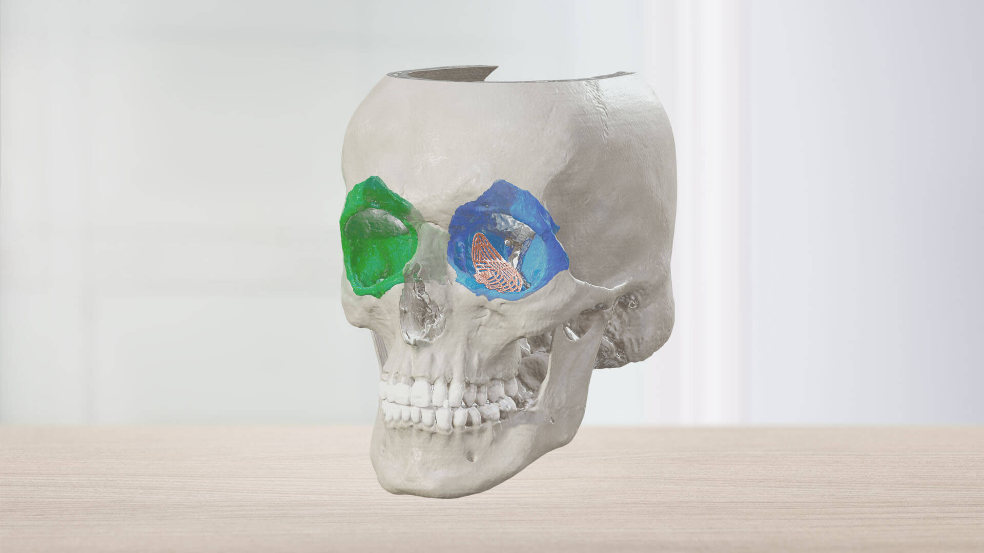
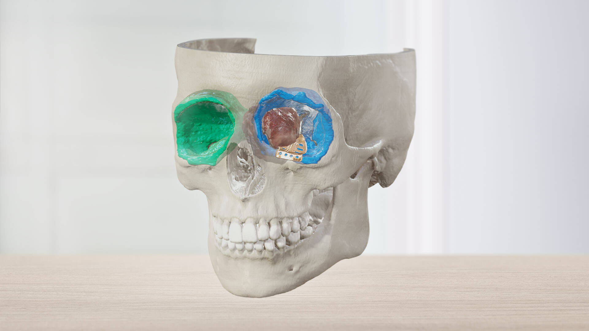
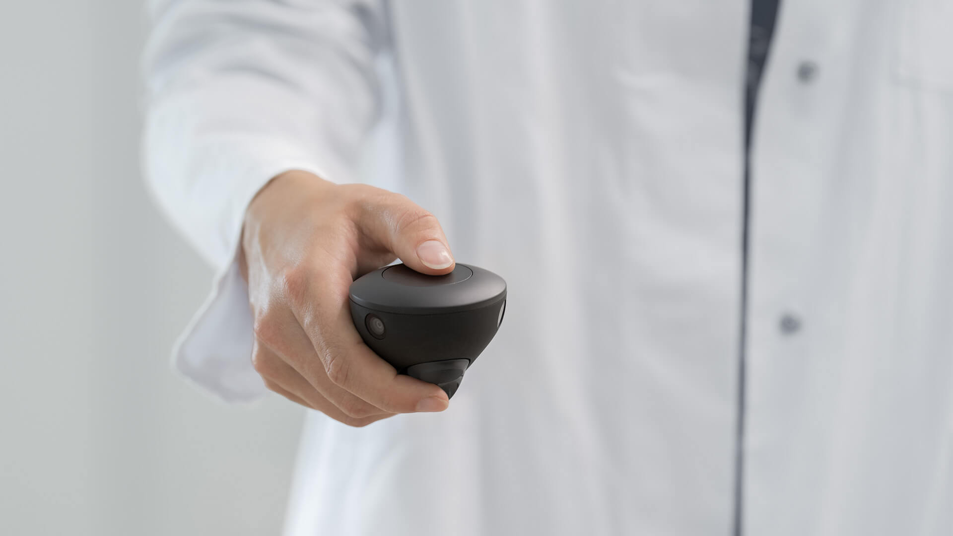
View key anatomical structures like never before. The Mixed Reality Viewer1 is a technology that works in tandem with Brainlab Elements to produce hyper-realistic 3D patient data. For craniofacial surgeries, the Mixed Reality Viewer can clearly display bony structures in an immersive environment while anatomical structures and implants important to procedures can be added selectively.
Enhanced reconstruction planning review and visualization in 3D
The Mixed Reality Viewer enables medical professionals to visualize and review complex reconstruction plans in 3D. With this technology, they can gain deeper insights into case-specific anatomy, spatial relationships and difficult-to-access anatomical segments like the optic nerve, various LeFort fractures and sphenoid sinuses.
Accessible, hands-on medical education
Medical students and residents have the opportunity to increase their understanding of patient anatomy and learn relevant surgical skills with the Mixed Reality Viewer. By offering features like multi-user sessions, instructors can also use the Mixed Reality Viewer to train students in an accessible, interactive and immersive 3D environment.
Immersive patient consultation
Medical professionals can use the Mixed Reality Viewer during patient consultation sessions to provide patients with a better understanding of the execution and potential outcomes of invasive CMF surgical procedures.

Our innovative surgical solutions are always just one click away
Explore the technical features of mixed reality for CMF cases
Leverage the potential of mixed reality and view the spatial relationships between bone, tumor and implant in 3D for craniomaxillofacial trauma fractures and tumor cases.
Experience our vision for mixed reality
Expand your perspective with the Mixed Reality Viewer
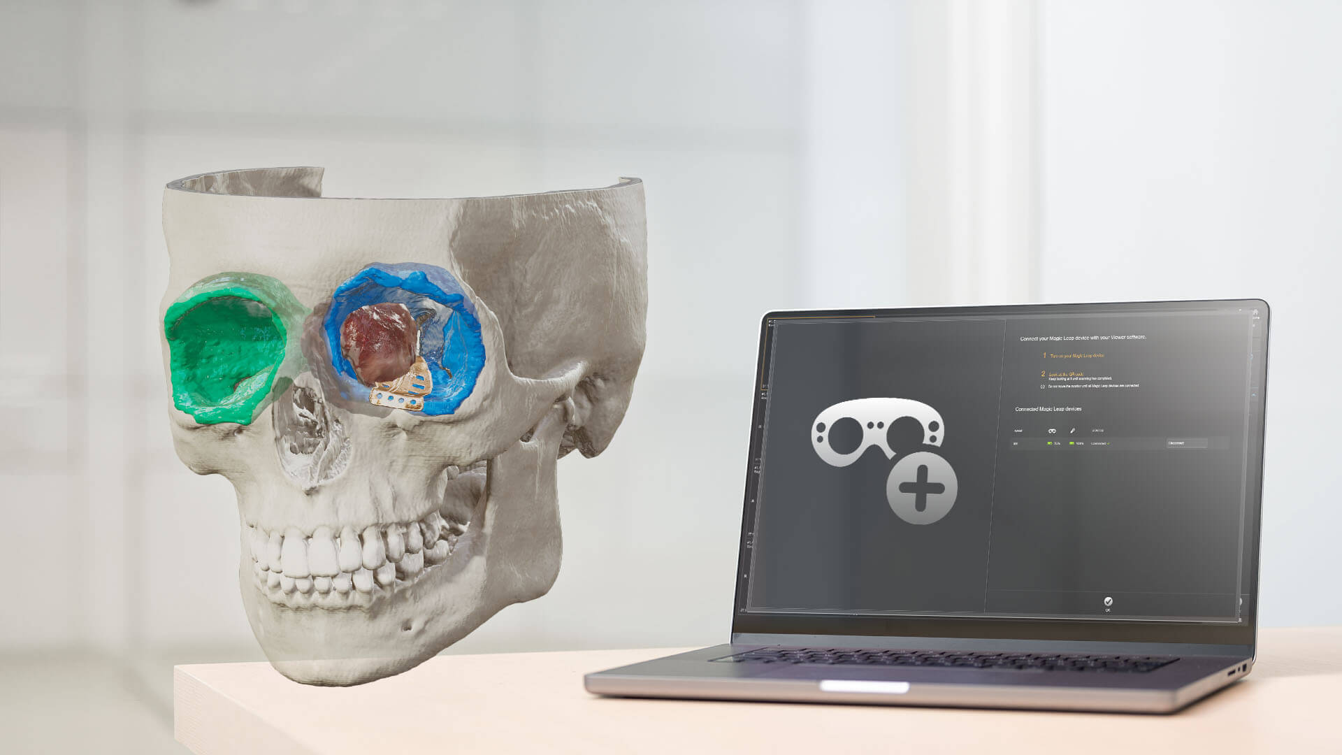
With a single click and a glance, your room is digitized for spatial computing. Images are transferred from the Elements Viewer software on screen into the room in front of you with the help of the Magic Leap spatial computing platform.


1. Start Elements Viewer
Access patient data from PACS and load it into the Elements Viewer

2. Boot your Magic Leap Device
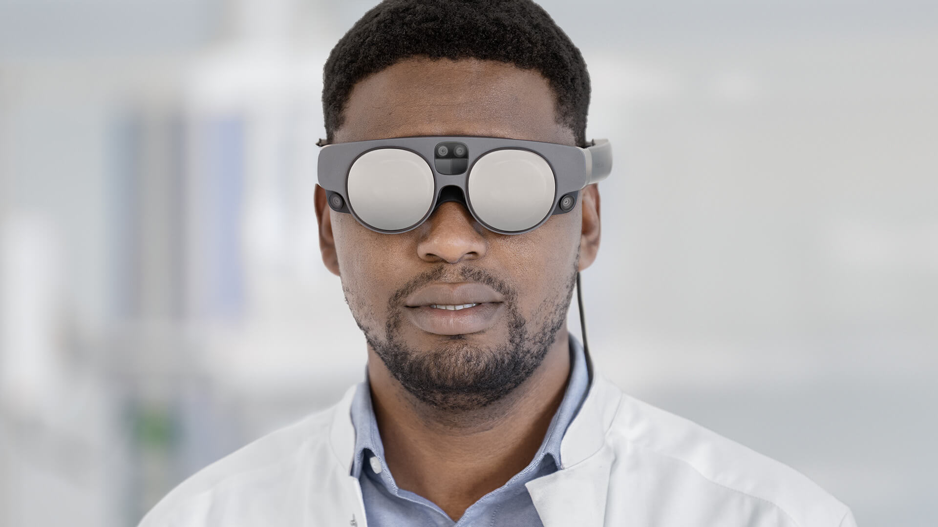
Boot your Magic Leap 2 headset and connect it to WiFi

3. Scan QR Code
Scan the QR code on the screen with your headset

4. Step into an immersive 3D model
Dive into patient data and interact with both virtual 2D images and 3D models
Not yet commercially available in several countries. Please contact your sales representative.
Discover our products.
Experience innovation.
Step into the future.
Experience innovation.
Step into the future.
Uncover the next generation of surgical solutions

