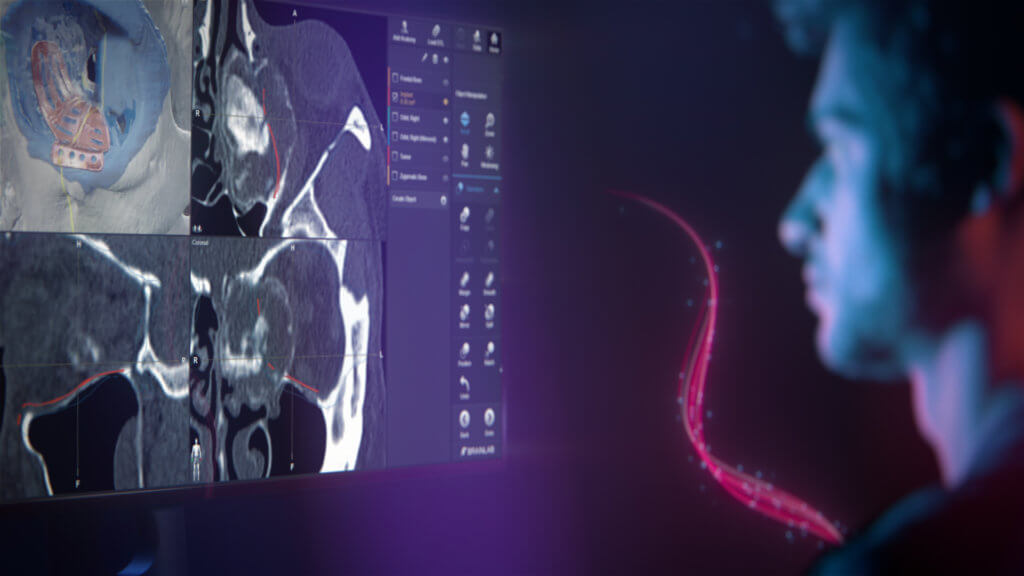
Header Visual: Brainlab Craniomaxillofacial Surgery Planning Software

Craniomaxillofacial PlanningUnique software tools
for unique cases

Discover more

Functionalities as individual as your cases

Craniomaxillofacial surgery planning is complex. With every case presenting unique challenges, you need tools that offer unique functionalities. Brainlab Craniomaxillofacial Planning software automates the steps that can slow you down while enriching patient data in new ways to help you make informed decisions. You can plan swiftly and intuitively, no matter how complex the case.

Take your plan a step further by adding mixed reality viewing, navigation1 and robotic intraoperative imaging to achieve your ideal outcomes for mid-facial reconstructions and tumor resections.

Automation at every step

We’ve built automation into every step of the planning workflow including data alignment,
image fusion, object outlining, segmentation and mirroring as well as implant positioning.
Save time while still thoroughly preparing for the procedure.

Customize your planning with Brainlab Elements

Brainlab Elements are software applications designed to automate and simplify planning and case preparation. Completely customizable, you can choose which Elements fit your reconstruction needs, from interactive data review to atlas-based segmentation and mirroring of structures to vendor-neutral import of binary STL files. Add the functionalities that will help you most, since these applications all fit seamlessly into your existing workflows.

Data Review

Easily align patient images

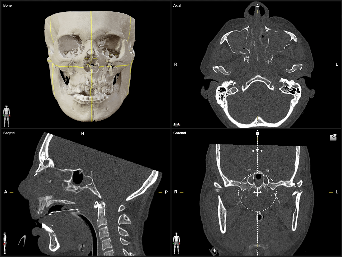
In CMF planning, correct alignment of the dataset is the first critical step. With Elements Viewer, imported DICOM data, such as MR, CT or cone beam CT scans, can be viewed in axial, coronal, sagittal or 3D and automatically aligned according to symmetry planes for simple comparison of both sides in one view.

Screenshot: Easily Align Patient Images for CMF Planning with Elements Viewer

Image Fuson

Easily align patient images

In CMF planning, correct alignment of the dataset is the first critical step. With Elements Viewer, imported DICOM data, such as MR, CT or cone beam CT scans, can be viewed in axial, coronal, sagittal or 3D and automatically aligned according to symmetry planes for simple comparison of both sides in one view.

Screenshot: Easily Align Patient Images for CMF Planning with Elements Viewer

3D Delineation

Easily align patient images

In CMF planning, correct alignment of the dataset is the first critical step. With Elements Viewer, imported DICOM data, such as MR, CT or cone beam CT scans, can be viewed in axial, coronal, sagittal or 3D and automatically aligned according to symmetry planes for simple comparison of both sides in one view.

Screenshot: Easily Align Patient Images for CMF Planning with Elements Viewer

Automatic Segmentation

Easily align patient images

In CMF planning, correct alignment of the dataset is the first critical step. With Elements Viewer, imported DICOM data, such as MR, CT or cone beam CT scans, can be viewed in axial, coronal, sagittal or 3D and automatically aligned according to symmetry planes for simple comparison of both sides in one view.

Screenshot: Easily Align Patient Images for CMF Planning with Elements Viewer

Virtual Reconstruction

Easily align patient images

In CMF planning, correct alignment of the dataset is the first critical step. With Elements Viewer, imported DICOM data, such as MR, CT or cone beam CT scans, can be viewed in axial, coronal, sagittal or 3D and automatically aligned according to symmetry planes for simple comparison of both sides in one view.

Screenshot: Easily Align Patient Images for CMF Planning with Elements Viewer

STL Import & Export

Easily align patient images

In CMF planning, correct alignment of the dataset is the first critical step. With Elements Viewer, imported DICOM data, such as MR, CT or cone beam CT scans, can be viewed in axial, coronal, sagittal or 3D and automatically aligned according to symmetry planes for simple comparison of both sides in one view.

Screenshot: Easily Align Patient Images for CMF Planning with Elements Viewer

What our customers are saying2

Brainlab Elements planning applications and mixed reality in practice

Ready to start planning?

Mixed RealityTake your plan from the screen into the room

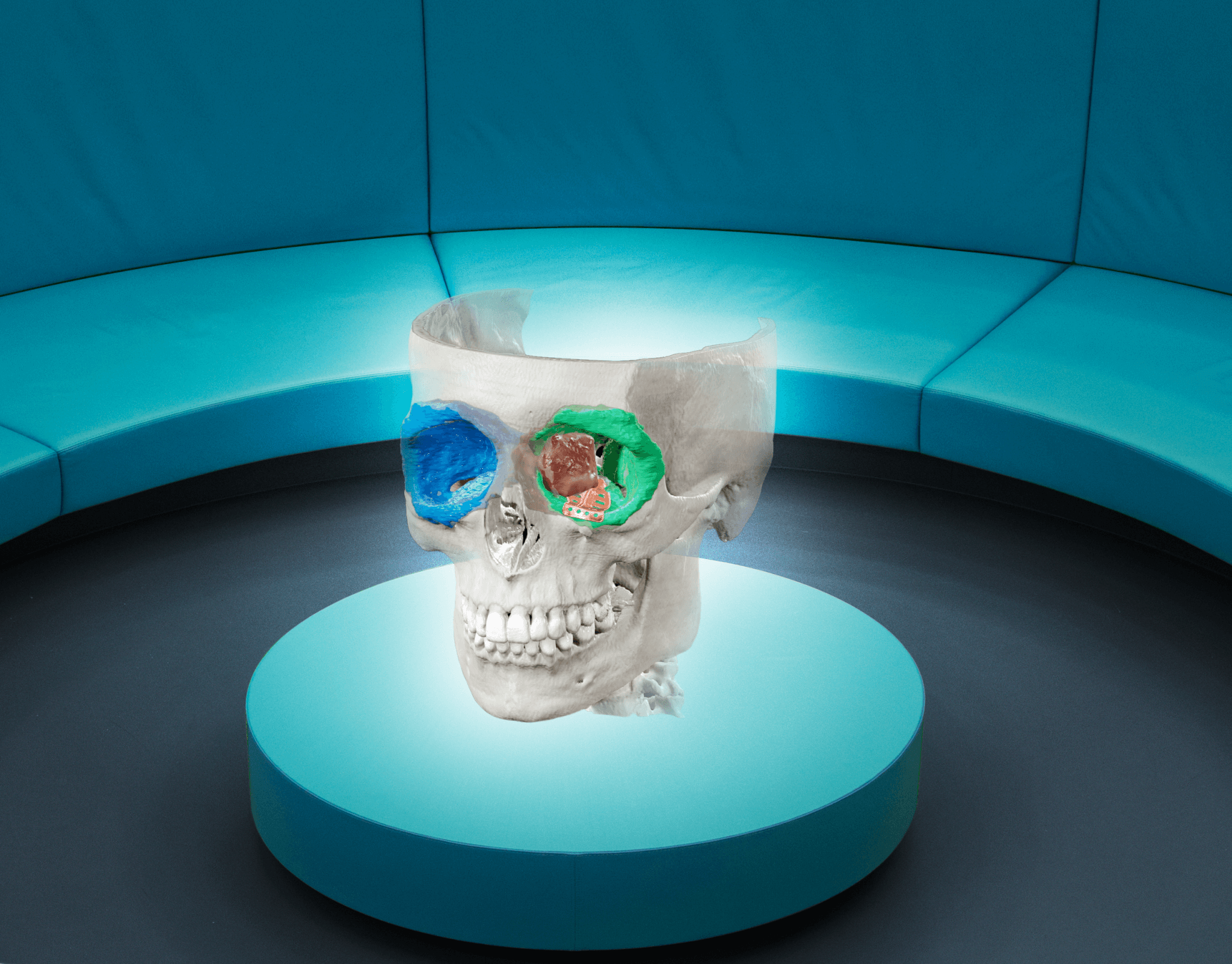
  • Mixed Reality frees viewing from traditional displays and turns the room into a virtual space to experience your patient-specific plan

  • Dive inside the patient’s anatomy in hyper-realistic 3D and view objects previously segmented with Brainlab Elements

  • Utilize the latest visualization technology for patient consultation and enable patients to better understand their diagnosis and the surgical approach

Cranial image viewed through Mixed Reality Viewer Glasses on a table in the center of the room

Learn more

Discover our collection of past CMF webinars that focus on the benefits of planning and mixed reality visualization. Be sure to register for our upcoming webinars!

  1. Not yet commercially available in several countries (CE mark available). Please contact your sales representative.

  2. The statements of the surgeons reflect only personal opinions and experiences and do not necessarily reflect the opinions of Brainlab or any affiliated institution.