250813_microscope_v022_01_16x9-2.jpg

Microscope Navigation for Cranial WorkflowsClarity in every layer

Surgical clarity where it counts. Microscope Navigation for cranial workflows brings patient data directly into view—supporting surgical accuracy through real-time, in-line visualization. Integrated augmented reality in the microscope’s optical path provides visual context to aid intraoperative decisions, adapt to anatomical changes and maintain procedural focus.

250915_microscopenavigation_cranial_benefit1_enhanceview.jpg

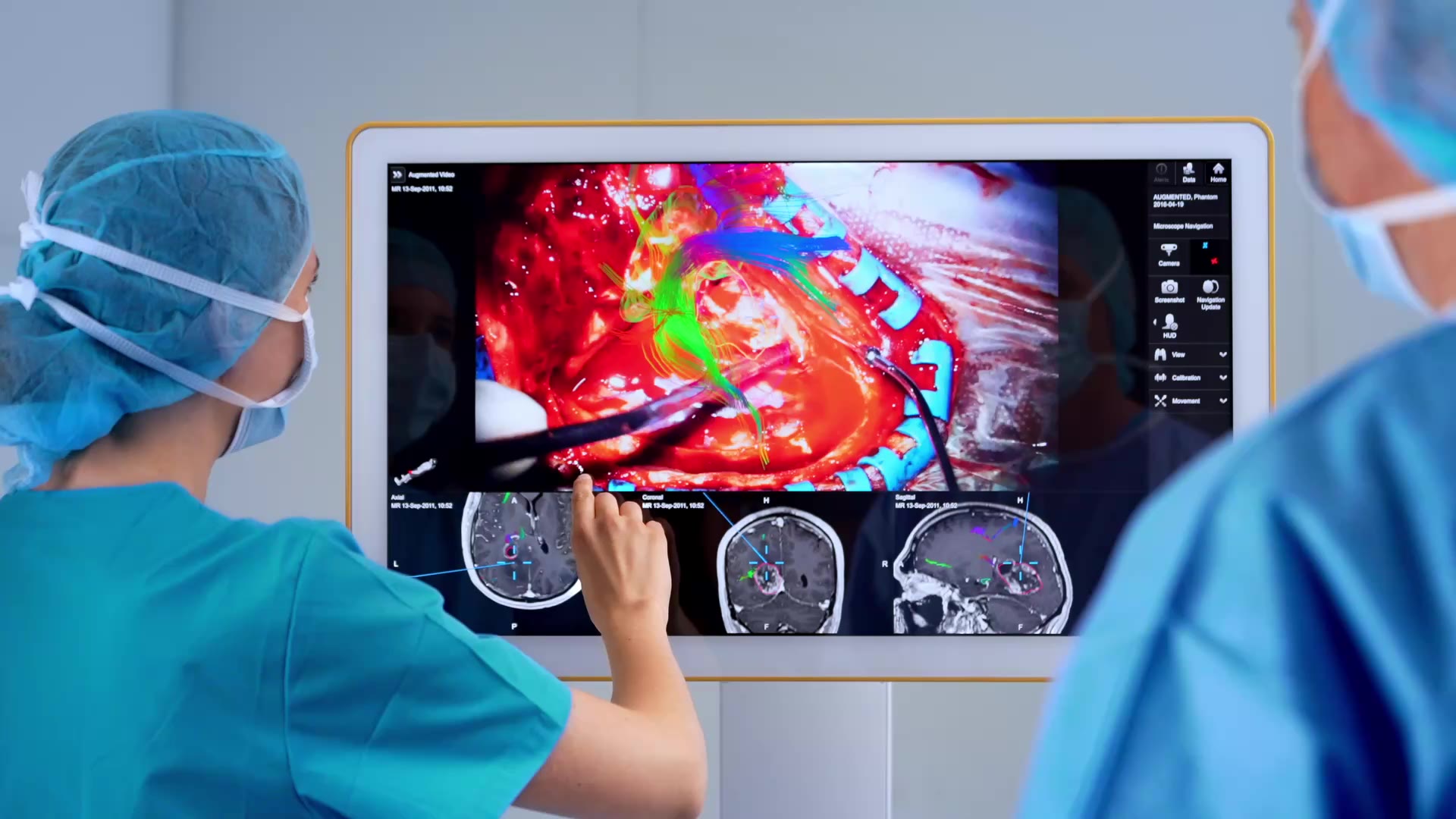
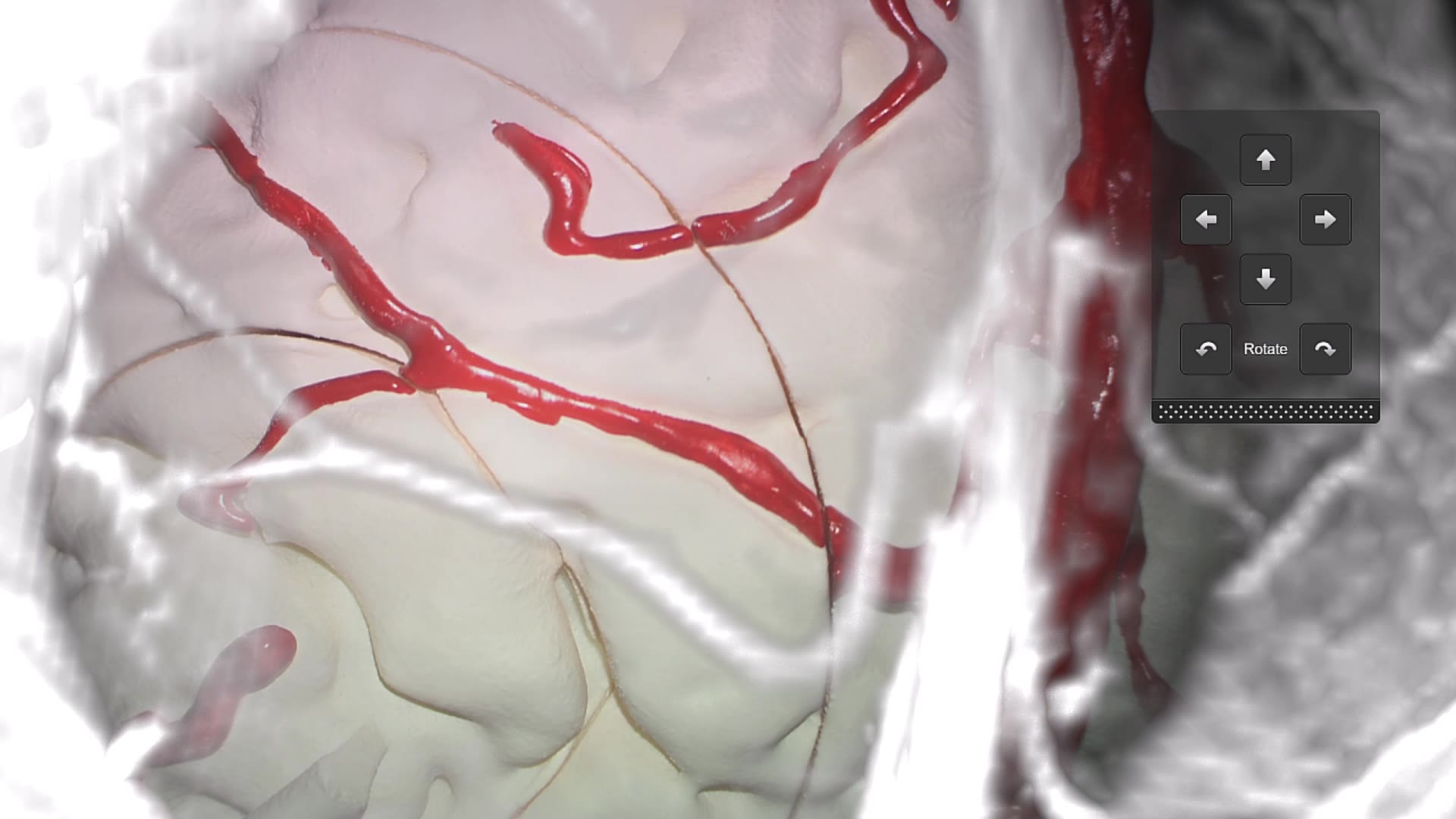
Enhance your view maintain focus

Overlay key structures like tumors, vessels or fiber tracts directly into the microscope view. Surgeons gain real-time spatial context without shifting focus, supporting confident decision-making throughout the procedure.

250915_microscopenavigation_cranial_benefit2_angleofapproach.jpg

Automate your angle of approach

Enable automatic microscope positioning with Microscope Navigation Robotic Movement. Align to anatomical targets or tools hands-free, enhancing ergonomic control, precision and efficiency with no manual repositioning required.

250915_microscopenavigation_cranial_benefit3_realignconfidence.jpg

Realign with confidence

Identify and correct small registration discrepancies by matching visible vessels to maximum intensity projection (MIP) images. Touch-based alignment enables fast, intraoperative updates—helping ensure navigation stays aligned with patient anatomy.

Learn more about our technology from clinical experts

Bring clarity to every critical moment

Augmented reality overlays and live anatomical alignment—directly in your microscope view. See how Microscope Navigation helps you stay focused, precise and informed.

Discover Microscope Navigation in the O.R.

Freedom of choice

Microscope use in the O.R. is evolving—and Brainlab adapts with it. Whether neurosurgeons prefer the traditional ocular view or the modern exoscopic approach with 3D video on large monitors, Microscope Navigation supports both workflows.
Exoscopic visualization helps improve ergonomics, reduce fatigue and support better posture during long cranial procedures. It also enhances team communication and training by allowing the entire surgical team to view the operative field in real time.

  • Auto-detection of integrated microscope reduces setup effort

  • Auto-detection of microscope reference array mounting position enables flexible surgical setup

250915_microscopenavigation_cranial_feature1_freedomofchoice.jpg

Intelligence in your line of sight

Microscope Navigation brings critical navigation data—like anatomical structures, planned trajectories and tool paths—into the live surgical view. Real-time AR overlays align with anatomy for continuous orientation without shifting focus to secondary screens.
By blending semi-transparent 3D volumes with the native view, Microscope Navigation supports spatial awareness and helps visualize targets and surrounding structures.
Visualization tools include:

  • Immediate Maximum Intensity Projection (MIP) for enhanced vascular detail

  • Overlay of eloquent fiber tracts to support surgical planning and resection

  • Interactive rotation of the microscope live video feed to explore underlying anatomy in 3D

cranial-microscope-navigation-feature-2-planerotation-mp4

Navigation updated

To support navigation accuracy after craniotomy, Microscope Navigation Update visualizes anatomical shift allowing the user to compare and match cortical vessels with their corresponding MIP overlays to update the registration.
Additional tools to support accurate alignment include:

  • Update patient registration during resection by intuitive, touch-based matching of planned landmarks to underlying anatomy

  • Repeat updates throughout the procedure, when required and feasible—also with any previously segmented objects

cranial-microscope-navigation-feature-3-navigation-update-mp4

Uninterrupted visualization—adapted to your workflow

Microscope Navigation brings essential information into the surgeon’s direct line of sight—on the navigation screen and in the microscope ocular. Adjustable transparency and a laser line indicating the focal plane help reduce visual overload and improve depth perception, supporting continuous focus and confident orientation.

  • Picture-in-Picture views with Zoom Lock preserve anatomical orientation at high magnification and allow flexible positioning

  • Visual control tools like transparency, fading and opacity adjustment improve clarity during delicate maneuvers

micr_nav_cranial_uniterruptedviewing_sr.jpg

Hands-free alignment with robotic control

Microscope Navigation for cranial procedures enables robotic positioning that’s ergonomic, efficient and intuitive. Instead of manual adjustments, surgeons can rely on autonomous alignment to any navigated instrument, planned trajectory or anatomical landmark—keeping focus where it matters most. 

Key capabilities include: 

  • Robotic alignment keeps the instrument tip centered and in focus, supporting efficiency and comfort during large-area resections 

  • Automatically maintains optimal angle and focus when following a navigated tool or adjusting the table, supporting uninterrupted workflow 

  • Eliminates manual repositioning, helping surgeons stay focused during complex, multi-angle resections 

cranial-microscope-navigation-alignement-robotic-control-mp4

Ergonomics meets precision

Exoscopic cranial navigation combines advanced visualization with ergonomic benefits, supporting surgeon comfort and enabling more collaborative workflows in complex procedures. It enhances both surgical precision and intraoperative communication.

  • Ergonomic design: Helps promote good posture and reduces fatigue—especially during long cranial surgeries—helping surgeons maintain focus

  • Shared visualization: Gives the entire team a real-time view of the operative field, improving communication, coordination and teaching

250915_microscopenavigation_cranial_feature6_ergonomicsprecision.jpg

Curious how this fits into your surgical workflow?

See the system in context—from planning to positioning and visualization. Our team will show you how Microscope Navigation for cranial integrates with your tools and techniques.

Use cases for Microscope Navigation

Microscope Navigation helps you stay focused on what matters—bringing patient data directly into view.

Focused precision for cranial tumor resection

Microscope Navigation supports tumor resection by delivering real-time, data-driven guidance directly into the surgeon’s line of sight. With in-line augmented reality integrated into the microscope optics, Microscope Navigation helps identify tumor boundaries, distinguish critical structures and adapt to brain shift—supporting safe resection while preserving healthy tissue.

250915_microscopenavigation_cranial_usecase_focusedprecision.jpg

Precise visualization for spine surgery

Microscope Navigation for spinal workflows supports a wide range of microsurgical procedures across the cervical, thoracic and lumbar regions. From degenerative cases such as TLIF and decompression for stenosis to complex spinal tumor resections, it provides precise guidance throughout your surgical workflow

250915_microscopenavigation_spine_usecaseimage.jpg

Explore more from the Brainlab Cranial portfolio

Let's talk about your O.R.

Whether you're refining cranial workflows or enhancing microscope precision, our specialists are here to help. Find out how navigation at the microscope could elevate your next case.

Proven impact in the O.R.

Additional Information

  1. Krieg S. et al., Preoperative motor mapping by navigated transcranial magnetic brain stimulation improves outcome for motor eloquent lesions. Neuro Oncol. First published online February 9, 2014.