Webinar

Computer-assisted Surgery for Reconstruction of Post-Traumatic Cranio-Orbital Deformity

Topics
画像誘導手術
CMF
手術ナビゲーション
手術計画
言語

English

Share this page

概要

Brainlab invites you to join our live webinar, “Computer-assisted surgery for reconstruction of post-traumatic cranio-orbital deformity”, on December 15, 2020 at 4:00 PM CET presented by Michael P. Grant, MD, PhD, FACS is Chief of Plastic, Reconstructive and Maxillofacial Surgery at the R Adams Cowley Shock Trauma Center, University of Maryland Medical Center, and Professor of Surgery and Ophthalmology, University of Maryland School of Medicine in Baltimore Maryland.

This webinar will cover topics including:

  • Learn to critically assess and diagnose post-traumatic defects of the frontal-orbital region
  • Understand the role of computer assisted surgical techniques in achieving optimal results
  • Learn to best evaluate the outcomes in these procedures, and avoid common complications


We look forward to meeting you online!

Language | English

In case you can not join the webinar, it will be recorded and shared afterward.

Participation is free of charge.

The views, information and opinions expressed within this presentation are from the speakers and do not necessarily represent those of Brainlab.

Speaker:

Michael P. Grant
Michael P. Grant

MD, PhD, FACS is Chief of Plastic, Reconstructive and Maxillofacial Surgery at the R Adams Cowley Shock Trauma Center, University of Maryland Medical Center, and Professor of Surgery and Ophthalmology, University of Maryland School of Medicine in Baltimore Maryland

Moderator:

Jana Neider
Jana Neider

Brainlab

Video Transcript

Jana: Welcome to our today’s CMF webinar and our webinar series for craniomaxillofacial surgery. My name is Jana Neider, and today we are live from my home office. As you might have heard, in Germany, we are again in lockdown, that’s why we are not today in the Brainlab office and today in the Brainlab Tower, but I’m happy to also have you here now from my home. So, it’s a pleasure for us that you are also joining today for the very exciting webinar on “Computer-assisted Surgery for Reconstruction of Post-Traumatic Cranio-Orbital Deformities.” Before I introduce you to our speaker, I would like to explain a few points. This webinar is live, but it will be recorded. Questions can be submitted through the online chat function and will be collected by myself and to be addressed to the speaker after he finishes with the lecture. As I said, the webinar will be recorded to be watched again at an independent time, independent from your attendance. For further questions, feel free to also use the chat function.

Now onto our speaker, Professor Michael Grant. Let me provide you with some of his details. Professor Michael Grant is chief of the Plastic and Reconstructive Surgery Center at the University of Maryland Medical Center in the United States in Baltimore. We are very happy to have Professor Michael Grant here today. We know him from a lot of different workshops that we had around the globe. We are very happy to have you here. And Professor Michael Grant is very much known for his intense knowledge on ophthalmology and plastic surgery, also of the reconstructive surgery, anesthetics as well as cosmetic ophthalmology surgery. Professor Michael Grant, I’m very happy that you are here today with us, and I have to say now the virtual stage is yours, and we are looking very much forward to your talk track.

Prof. Grant: Well, thank you, Jana. Thank you for your team for organizing this.

Jana: Thank you.

Prof. Grant: All righty. I think we should be ready to go.

Jana: Yes, we are ready to go. We can see and hear you, and we also do see your slides.

Prof. Grant: Great. Well, thank you very much again for inviting me to give this talk. I think cranio-orbital deformities and cranio-orbital trauma give a good chance for us to talk about the digital workflow that would help give us the most reproducible and safe results for our patients. So, when we talk about a computer-assisted surgery for reconstruction of these things, we’re going to go through sort of a workflow, and I want to explain the various parts of it to you. I am head of plastic surgery at the R Adams Cowley Shock Trauma Center, which you see here. It’s a large self-contained adult trauma center in Maryland. We treat about 8,000 to 10,000 seriously injured patients per year. And you see our building there on the left.

When we think about cranio-orbital trauma, we’re really talking about frontal orbital trauma. We’re talking about injuries that involve the orbit and the frontal bone, as you see illustrated here with this patient with an obvious injury to both orbits and the central frontal bone. The frontal bone contains the frontal sinus. And I don’t really want to talk at length about management of frontal sinus injuries. That in and of itself is something that we could talk for an hour about. But most of the patients that I’m going to show you here today, in fact, I think all of them have injuries to their frontal sinus, which required management. And we’ll talk a little bit about the management that was required in the setting of those patient examples that I’m going to give you, but just a few terms to sort of talk about here by way of introduction. The anterior table is the outer portion of the frontal sinus. The posterior table is what separates the back wall of the frontal sinus from the brain, and the naso-frontal ducks, which you see here in this patient, which allow the frontal sinus to drain into the nose.

Management of the frontal sinus depends on injuries and the integrity of those three structures. So, we’ll talk a little bit about those structures as we go along and look at our patient examples. So, what about the orbit? What do we need to consider in treating the orbital part of an orbital frontal injury? Well, the two main problems that we see in these injuries are globe malposition and double vision. And here are two patients that have both of those problems, globe malposition, and double vision. You can see the patient on the left has an obvious depression of her globe. Her left globe is lower than her right. She has double vision. You can see on the right side this patient has a globe that’s higher on the right, which is the injured side than the left.

So, getting the globe in the right position is critical, and computer-assisted techniques can really help us with getting that reproducibly correct. We know a little bit about the relationship between volume and position of the eye, that is whether or not the patient has enophthalmos. So, we know that for about one square centimeter or one CC expansion in orbital volume produces about a one-millimeter decrease in the axial projection of the eye, or what we call enophthalmos. So, the relationship between volume and eye position is pretty well understood. What’s not as well understood is the relationship between volume and I would say shape and function. And we’ll talk a little bit about that as we go along.

Why would changes in volume and shape change function of the eye? Well, if you think about the structure of the orbit in the untraumatized state, there’s a drawing here on the right and a silver stain on the left from a gentleman named Koornneef. Koornneef was a Dutch ophthalmologist and anonymous, and he described this really intricate microstructure of the internal orbit. And what you can see from this is that the orbit is organized by these septae, which he cleverly named after himself, called the Fibrous Septae of Koornneef. And you can see the eye muscles are arranged radially and they travel in these little sleeves, and these little septae organize all of these structures in the orbit.

When an orbit is traumatized, you see a CT scan on the left here, just a 2D view and Koornneef’s description in diagrammatic form of what he thinks happens. And you can see this is long before CT scanning, and you can see that he argues that this structure is disrupted, that some of the fat here that’s intraconal becomes extraconal and this nice microarchitecture is arranged, and you see exactly that going on here on the left. So, I don’t think it’s a huge leap of faith to think that if you allow something to heal like this, that it might not work as well. So, that’s why accurately reconstructing the inner orbit is important in function, and that’s how computer-assisted surgery can help us.

So, as we go along through this in patients that I show you that have double vision, we want to think about is double vision a surgical problem? Because I would argue that it is, and I want to talk a little bit about functionally rehabilitating people. We’ve gotten very good at putting bones back together very accurately. And I’ll show you a study that we’ve done that demonstrates that, but we really want to make people functionally better, and how can computer-assisted techniques help us with that?

So, the third term to define here, we talked about cranio-orbital, so cranio meaning, in this talk, really frontal bone injuries. Orbital, we’ve talked about what’s important in that. How does computer-assisted surgery figure into this? And when we think about computer-assisted surgery, what I want you to think about is a digital workflow, and what does that look like? Well, here’s a diagram just from Brainlab of how they conceptualize a digital workflow. And it starts with planning and analysis in step one, then moves intraoperatively to navigation intraoperatively, then moves to imaging, which is either intraoperative or post-operative but is required. So, really, imaging is central in the workflow.

So, I think we need to move away from this concept that we don’t image patients after facial reconstruction. And there are still places that don’t do this routinely. And it’s obvious, I think, if you look at the workflow that imaging is a central role in the workflow. And then that imaging brings us back to analysis and planning again, post-operative quality control. We can look at our scans and we can fuse them with the plan and we can figure out how well we’ve actually done in executing the plan. These datasets can be exported as you see here, and that can be done for biopsy landmarks, they can be done to make STL models, it can be done to make patient-specific implants.

We’ll talk about that as we go along today, but the planning is what generates these datasets. You see in the middle here reference to mixed reality. So, some of these datasets, we can then manipulate in a mixed reality typesetting. And there was a webinar about a month ago on that that showed the power of mixed reality and augmented reality in digital workflow. And I’m not really going to talk about that, but just suffice it to say that those techniques are out there.

So, let’s really drill down and look at what is computer-assisted surgery? It’s not computers doing operations. Patients often ask me that, “Well, is a computer doing the operation?” No, it’s a workflow. So, we have a clinical situation, an injury, a tumor, something. We image the patient so we have our dataset. Imaging takes us to planning. So, from there, we can visualize, we can diagnose the problem, we can look at the fracture pattern. We can decide what sort of an injury it is, and that comes from our 2D and 3D analysis, and then we can move to generating a template for reconstruction. So, that’s going to involve segmentation of the dataset, mirroring, maybe some simulated surgery or virtual surgery, which I’ll show you some examples of as we move forward. And then that’s going to produce a virtual model or template, and that’s what we’re going to use for our reconstruction. That virtual model or template can be taken virtually to the OR, loaded into our navigation system, and then we use that dataset to guide our intraoperative surgery. So, that’s how intraoperative navigation fits into this.

Notice that intraoperative navigation here, which I think is what a lot of people assume that when one speaks of computer-assisted surgery and Brainlab, they sort of focus on the navigation. Navigation is really a small part of what we do here, a very valuable part, but a small part of the overall digital workflow. So, that surgery produces some result, and that takes us back to clinical evaluation of the patient. What do we have? Have we fixed the problem adequately? That’s going to require imaging, so we’re going to do an intraoperative or a post-operative CT depending on how your unit is equipped, and that’s going to take us back to analysis again, post-operative quality control, fusion, and then follow-up and continued management of the patient. So, I borrowed this slide from Nils Gellrich who has taught me a lot about digital workflow and application of these technologies to cranio-orbital defects. And I think it’s a really nice way of illustrating the power of digital workflow in taking care of patients.

So, a logical question to ask is, does all this work make a difference? Does this sort of analysis and planning help us in achieving better results? So, Nils was really the catalyst for this study that was completed a few years ago, called the Orbita 3 study. You can see several of the speakers that have spoken in this forum through the Brainlab series are included, Nils, Alexander Schramm, Frank Wilde, Rudiger Zimmerer, me, Majeed Rana. All are people that have adopted these principles and this application of a digital workflow.

So, in this series of studies, what we did is we looked at if one takes a prefabricated implant and just places it based on clinical knowledge, or one takes an implant that has been manipulated maybe from an STL model or something like that and places it with computer-assisted surgical techniques, meaning planning and navigation, and compare that to what happens if you take a patient-specific implant and place it with guidance. And you can see that it is much more accurate as we go from basically freehand placement of a prefabricated implant to computer-assisted placement of a patient-specific implant. And you can see that the orbital volume we achieve in using that technique is much better if we use this digital workflow. So, let’s talk about some patients’ examples and see how we apply this workflow to patients with cranio-orbital defects. And I’m going to go from low-velocity defects to high-velocity defects and show you how this can be applied in any of these clinical situations.

So, here’s a gentleman that was hit in the left side of his face at work by a piece of metal that flew up and hit him in the left side of his face. You can see this is an acute injury. He was injured about three days ago. Here are the 2D views showing damage to the anterior table of his frontal sinus and a two-wall defect of his internal orbit. You see his medial wall is fractured here, and his orbital floor is fractured, and the transition zone is fractured.

So, we can take this patient’s dataset and we can mirror the unaffected right side to the left side, and that becomes our template for reconstruction. And you can see here in red the template showing us where the damage is and providing guidance for accurate reconstruction. We can then virtually place a prefabricated mesh preoperatively, check to make sure that it fits what the right orientation is as we’ve done here, and then we can download that as a dataset, take it to the operating room and use it to navigate to assure that we’ve positioned the implant correctly.

Once we’ve done that, we can do a CT scan intraoperatively as we’ve done here. And that shows the overlying plan, the implant in place, and the patient’s bony reconstruction. And here he is post-operatively. One of the things that this sort of analysis allows us to do is that allows us to use minimally invasive techniques. So, this was all done through a combination of a transconjunctival incision and an upper eyelid crease incision. So, no coronal incision, no significant exposure, but because we have accurate imaging, because we have accurate planning, we can plan small incisions, and that allows us to do more through a smaller exposure.

Let’s turn now to a little higher velocity injury. This is a young patient who was involved in a high-velocity motor vehicle collision. She was unrestrained and hit the steering wheel and windshield of the car. She’s brought in. You see her here. Brought into our trauma center. She has a laceration over the dorsum of the nose that’s been repaired. You can see the classic sort of deformity of this frontal orbital injury retrusion of the radix of the nose, depression, upturning of the dorsum and tip, and you can see that she has fairly significant ecchymosis on the left side of her face. Here are the 3D views of that CT scan. So, you can see that she has this damage to the frontal bandeau and a Type 1 nasal-orbital-ethmoidal injury, and an internal orbital fracture seen here. So, the patient was cleared by neurosurgery. We took her to the operating room, and we laid her down for surgery.

And as I was taking a look at her after I removed the sutures, we see this clear fluid pooling. And if you dab that clear fluid away, it pulls again, and if you dab it away it pools again. And what this is is cerebrospinal fluid. So, this patient, by definition, now has an injury to her anterior cranial base and a dural leak, and now she needs a different operation to fix that, then just an operation on her orbit and NOE. So, with my neurosurgical colleague, we did a coronal dissection, raised a pericranial flap, did a frontal craniotomy, and sealed off her anterior skull base, and fixed the outer frame of that NOE injury. We did not want to do any intraorbital dissection because the damage to her cranial base is right up in this region, and I would have to dissect up in there to fix her orbital fracture. So, we reduced the outer frame, addressed the CSF leak, and then decided to treat her orbit secondarily.

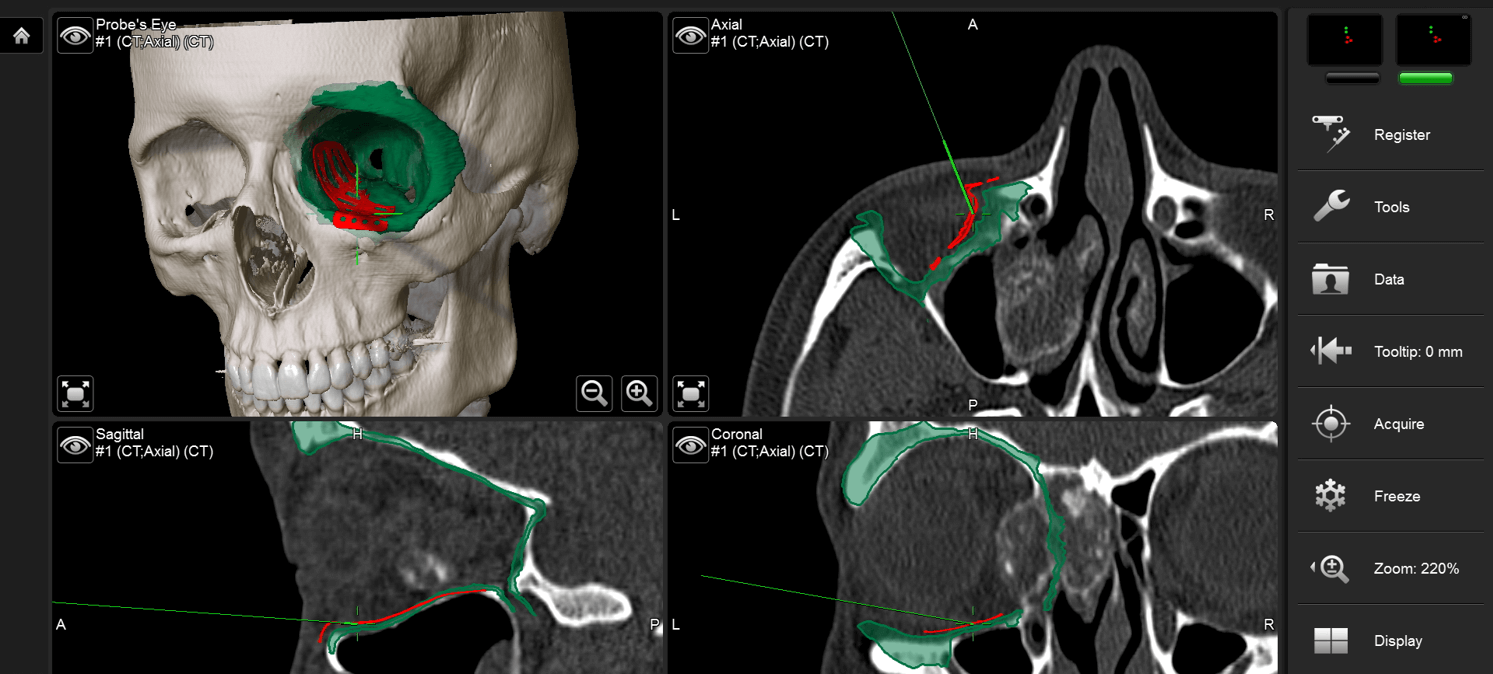
So, here she is now about a month after. You can see she has obvious enophthalmos now with that large two-wall fracture. Here is her unaffected right side and her left side. She has double vision in all fields of gaze. So, she now gets her dataset exported to an industrial partner who makes a patient-specific orbital implant. And you can see the orbital implant that we’ve selected here. So, I’m going to use this implant to reconstruct her internal orbit and stabilize the lateral portion of that NOE fragment. You can see here. So, I’m going to reduce that. I already have a plate here that I placed in the previous operation, and you just see the plate there in BeneView. And then I can virtually position that implant as you see here in the STL file, and I can then navigate during the surgery, and I can assure that I’ve positioned that plate correctly.

Now, remember the CT is not real-time here. The navigation is real-time. So, in the red, I’m tracing along this implant, and I can verify that it’s in the correct position and that I’ve adequately restored the integrity of the internal orbit. And you can see just dissecting here posteriorly to the posterior ledge to make sure that the implant is positioned appropriately. And then here is her intraoperative CT scan with the implant in place, and here she is about four months after surgery with no double vision and resolution of her enophthalmos, and fortunately, her CSF leak as well.

Let’s talk now about a little higher velocity injury. Here’s a patient that was walking along a motorway because she had difficulty with her car. She got out of her car and was hit in the left side of her face by a mirror from a truck. She had a significant head injury, subarachnoid, subdural hemorrhage. Went for an emergency craniotomy and reconstruction, all at an outside hospital, and she was then referred to me with this difficulty with the physician of her left orbit. You can see her left orbit is down. Her eye is forced down and out. She has misalignment of her eye because her eye is being pushed so far laterally and down that she can’t adequately fuse the two images. So, she has significant double vision. Here’s a 2D view of her reconstruction.

See, she has this PEEK implant that’s been positioned here, and I’ll show you a better image of that in a second. She had a significant CSF leak, so some sort of graft was placed in her cranially to try and seal her anterior skull base. But you can see just from this 2D image that her eye is being pushed down and out in this direction. She has an untreated floor defect here, and a smaller medial wall defect you see right here. So, basically, her eye is being pushed down by this distorted orbital roof and frontal bone repair into this untreated defect in her floor. So, you may just say, “Well, why not just fix the floor and push her eye back up?” But you can see in this view that there’s nowhere for the eye to go. So, to reconstruct her floor, we have to reconstruct her roof, which means we’ve got to do something with this piece of bone.

So, here’s just a couple of 2D images looking at mirroring the right side of her orbit and trying to figure out where that orbit needs to be to get her eye in the correct position. And you can see that this bone that’s been pushed down in her orbit we’re going to have to deal with to get her eye back into better position. So, here’s a model showing the problem. Here in white is this piece of PEEK that was made at the outside hospital and placed over this frontal bone defect and temporal bone defect, which you see here and here, and back in here is where this bone is intracranially.

So, working with my neurosurgical colleague, what we’re planning is to reopen her coronal incision, remove this piece of PEEK. That will allow us exposure to the anterior skull base, and then we’ll remove those pieces of bone, repatch any dural tears, and hopefully, allow her eye to come back up into position. So, here’s our plan. You can see in red are the two pieces of bone that need to be removed. This is our planned implant that we designed for the roof. Here, she is intraoperatively after positioning of this roof implant and floor medial wall implant. Remember, you can’t see the PEEK radiographically, and here she is post-operatively with her eye now in better position and now able to fuse and achieve a single vision versus the double vision that she had had since the accident.

Little higher velocity injury. Now, this is a ballistic injury to the face, specifically the right and left orbit frontal bone, roofs of both orbits that you see here. So, this is a victim of the Las Vegas gunshot or shooting massacre that took place a little over three years ago. She was evaluated and treated on scene in Las Vegas and evacuated to our institution about five or six days post-injury. So, she’s had a decompressive craniotomy. You see the staples are in place here. You can see this devitalized and damaged frontal bone, all these fragments of bone and ballistic material. She’s lost her right eye and this material draining down her cheek, the cerebrospinal fluid, so she has a large dural injury and dural leak.

We took her to the operating room with our neurosurgical colleagues and turned down the coronal flap. There’s this piece of stable bone that you see right here. You’re looking at this piece of bone from above. This is a big piece of dural gel which we reinforced and fixed the leak in the anterior skull base and then planned for orbital reconstruction. So, because both orbits are damaged here, there isn’t anything to mirror. So, by importing standard skulls and trying to match shape and volume with the intact orbit, we were able to come up with our ideal orbital shape and position. So you see on the right, we need a roof implant, a floor, and medial wall, and on the left, we need a roof and medial wall. Now, important in this is that she doesn’t have a right globe, only a left. So, she’s monocular. So, we have to take extreme caution in reconstructing the left side because of her monocular status. You might say, “Why not just leave it alone?” Well, if we don’t repair the left roof and medial wall, her brain is going to herniate into her orbit and threaten the vision of her left eye.

So, here are our planned implants. You can see the three wall reconstruction that we talked about on the right, the two wall reconstruction we talked about on the left. For the frontal bone reconstruction, because this was contaminated and her frontal sinus was involved and had to be obliterated, we decided against an alloplast and used split cranial bone to fix her frontal bone. And Dr. Groves, the neurosurgeon, Dr. [inaudible 00:30:51], and I performed this secondary reconstruction of her skull base and bilateral orbits and dural. So, you see here an intraoperative view showing the roof implant going in on the right, and now on the left here is the split cranial bone going on for her frontal bone, and here she is on the table, intraoperative imaging showing the positioning of the implants in the orbit on the right and on the left. You can see all of this material in here had to be debrided out to get those implants in position.

Here she is acutely after stage one. So now we designed some onlays to try and improve the contour of her forehead. Here she is after that’s been allowed to heal. Here you see those little irregularities in the frontal bone graft, here with some onlay with porous polyethylene. Now, here she is after her second stage operation, and here she is now after fixing her eyelid and getting her prosthesis. So, you can see we go from the picture on the right to the picture on the left showing reconstruction of her frontal orbital defect.

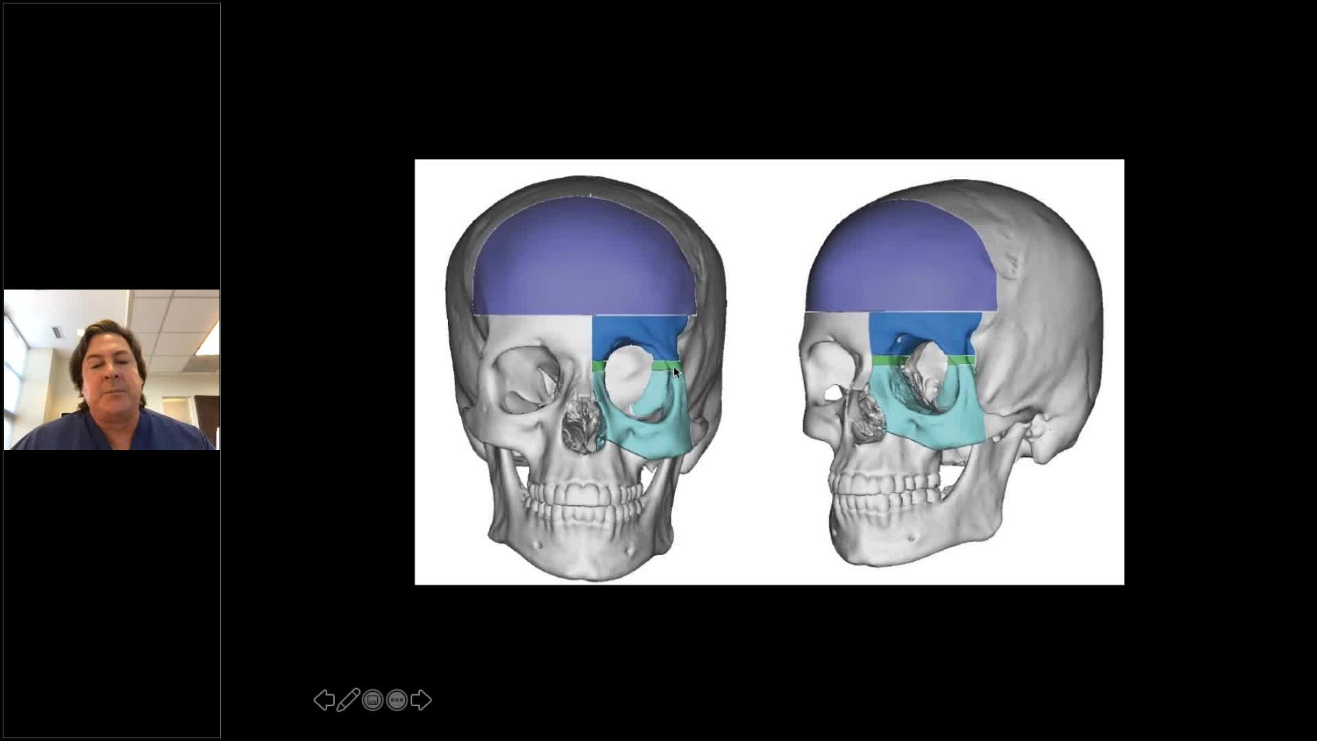
So, I’ve shown you a number of traumatic injuries that we can apply this digital workflow to. Can we apply it to other problems? And the answer is, of course, yes. Here is an example of how we’ve applied this workflow to quiet problems that are nontraumatic. This is a young woman who had had multiple operations for neurofibromatosis affecting her left orbit. And you can see that her eye is down when compared to the right side. It’s down, and it’s deviated a little bit medially. So, we can use this same digital workflow to perform a virtual box osteotomy, the classic Tessier box osteotomy. So, we’re going to do a frontal craniotomy. We’re going to take the superior portion of her orbit because you can see this orbit is like a big oval.

So, conceptually, the frontal bone has to come down and the inferior orbit and zygoma needs to come up. So, what we have to do is decide how much to do that, and by doing that, we measure the volume and shape of the right orbit, transpose it to the left. We have about one centimeter of bone that needs to come out here, one point two centimeters of bone that needs to come out so that we can move this bone down and move this bone up. So, all that can be done virtually, and you can see we’ve done the virtual box osteotomy here, and now we’ve designed patient-specific implants from that dataset to be positioned once the box osteotomy is performed.

So, using cutting guides, we’ve cut the box, this is all virtually now, and design these implants. And then we can take that plan to the operating room virtually, perform the operation, position the implants, verify that with navigation, and then ultimately with CT scanning. So, we have done the procedure here. Here are the implants in place. In yellow, you see our predicted volume, in orange, you see our achieved volume. So, virtually, 100% correlation there. And here she is after just one operation. Here she is at about three or four weeks. Here she is at about three months. All of these patients need eyelid surgery because their eyelids have been stretched. So, she’s going to go on to have a lateral canthoplasty and ptosis repair once the swelling settles out. But this is a young lady that has had a dozen operations to try and fix this problem, and in one operation we’re able to achieve pretty good symmetry by using this digital workflow. And here’s just a reminder of where we started, so you can see much better positioning of the globe or double vision has resolved. And just as I mentioned, with some eyelid surgery, we can make her quite symmetric.

So, I just would like to alert you to a study that we just performed at our institution here, Selim Gebran and Joey Lopez, my research fellow and a former chief resident here have really done an excellent job looking at visual outcomes and these frontal orbital injuries. And this is an article that’s just going to press now in plastic and reconstructive surgery. If you’re treating traumatic injuries of this area, this is something you may want to put on your radar screen. It sort of gives you our algorithm in how to treat roof and frontal orbital injuries in adult patients.

So, just to finish up here, what are I think the keys for success, at least from my perspective, in treating these patients and employing this sort of workflow? So, prompt, precise treatment of frontal orbital fractures based on preoperative evaluation and planning. So, that’s step one of that digital workflow. It’s probably the most important step is that analysis and planning that goes in upfront. Once we have the plan, we employ our digital workflow. In other words, we take that plan, we take it to the OR. In most cases, that then translates into intraoperative navigation. Virtually, all of our patients go down that pathway. It’s really only the simplest cases that we don’t do navigation for these days. And then intraoperative or post-operative imaging and quality control in all cases. That’s how you verify that you’ve executed your plan. That’s how you get reproducible and safe results with these complicated injuries for your patients.

I’d like to thank Nils Gellrich, Professor Gellrich from Hanover who provided a couple of slides, as I mentioned, for this talk. Nils came from Freiburg where he was the vice-chair in that department under Reynard [inaudible 00:38:28] who really has spawned a whole group of people that have pushed the envelope in this. Alexander Schramm who then went to Hanover with Nils, and then many of their trainees and collaborators that I mentioned when reviewing the Orbita 3 study have gone on to help define this digital workflow for craniomaxillofacial surgery. So, I’d like to thank our colleagues at Brainlab for allowing us to present this talk and thank them for partnering with us to employ these technologies to help our patients. And with that, I’d be certainly happy to take any questions.

[00:39:15]

[silence]

[00:39:30]

Jana: Thank you, Professor Grant, for the excellent presentation. It’s just amazing cases that you showed, especially the one from the gunshot. That was really impressive to see. And we already received a lot of questions for you. I will address them in a second. So, just to remind everyone that is attending the call, you can still send us questions using the chat function, and I will still address them in this question and answer session now with Michael. So, Michael, the first question to you, and it actually comes as a form of thank you for you, so thank you for the wonderful presentation. You showed a lot of interesting cases. How do you actually treat bilateral defects? I think you also answered that question in your second part of the webinar, but maybe you can just summarize it again because maybe the person just joined later.

Prof. Grant: Yes. So, that’s a great question. Bilateral defects are more challenging because there isn’t a contralateral control that we can mirror and use for our template. But we are able to import standard unfractured datasets and make comparisons to try and find a patient that has a similar volume and shaped orbit using the unfractured parts of the orbit and mid-face that are present as keys. Also, some of our industrial partners that make implants have libraries of standard skulls which they can assist you with in trying to analyze these defects and help you with the preoperative planning. Those companies that will do that for you will send you with the implants an STL file that you can upload directly into Brainlab and use for the intraoperative navigation piece. So, it’s a way to partner with other companies such as KLS Martin, DePuy Synthes, which will make these patient-specific implants for you and that you can use then in this same platform that I illustrated during my talk.

Jana: Thank you, Michael. I think you highlighted a very important point. So, Brainlab is a completely open platform that is working with a lot of independent implant vendors, so we don’t limit our customers to work together with one or the other. So, you can always decide what is the best implant vendor for your choice? Especially for very specific cases. You might even change the vendors. So, we are completely open to collaborate with all of these.

Prof. Grant: Yes.

Jana: The next… Thank you. The next question, Michael. I recently saw the “Mixed Reality” webinar with Professor Strong, how you can imagine to use mixed reality for planning and treatment of complex CMF cases. Is there something you could imagine, and is it also something interesting for surgical procedures?

Prof. Grant: Yeah. So, we’ve played with that system a little bit, and I can definitely see it as a way, for example, in a group setting with a group of trainees to look at a plan or devise a plan where we can all see and manipulate that plan sort of together. When we do planning now, it’s usually two or three of us sort of huddled around a workstation looking at a 2D screen, and that works fine, but in mixed reality, you’re able to experience the three-dimensional flavor of the plan and the injury and what you’re trying to treat. So, I see it as sort of the next step in a way to make planning more interactive. And I think a lot of us think and see things three-dimensionally but we’re forced into 2D platforms. And I’ve noticed with my residents just using the planning aspects in Brainlab now without the mixed reality just how beneficial it is for them to see things three-dimensionally before trying to tackle those problems in the operating room.

Jana: Thanks a lot, Michael. The next question was…I think we got it, one of the orbital cases that you showed, and the question was, how do you measure actually the orbital volume? Is this something that you can do also within that software, or you have to have specific other software for that?

Prof. Grant: So, you’re able to measure orbital volumes in the Brainlab system, and also again, industrial partners that give that design implants for you can help you measure orbital volumes. The challenge in measuring orbital volumes is deciding where you define the front of the orbit. So, some people define the front of the orbit just as a tangent line, example from the frontal bone to the inferior orbital rim. And looking at that volume, some people define it as the soft tissue outline on a surface map of a CT scan. And there are a number of other systems out there that will measure orbital volumes for you. I guess I don’t get really hung up on the actual measurement as much as I do get drilled down on the real shape and volume in terms of the implant. I know that if I mirror the contralateral side and use that for my template, that I have essentially treated the orbital volume appropriately. Mark Metzker and others did a number of studies showing there’s very little difference between volumes of the right side versus the left and vice versa. So, the actual number that we put on it I don’t think is important as the process.

Jana: Thanks a lot, Michael. The next question is, actually, who is doing the planning? Is it you, is it a team approach? Do you need us or someone from Brainlab that helps you with the planning? Yeah. So, that was the question. Maybe you can explain a bit more how the planning itself works.

Prof. Grant: So, the planning is typically done by me or one of our fellows or residents. The residents really like the planning because a lot of them are very sort of tech-oriented and they find it fun. The software is basically menu-driven. So, once you’ve planned two or three cases, it’s pretty straightforward to load the dataset, to align the dataset, then mirror the injury to generate your template. And then, you know, the real fun they also have is then going back with the patient’s intraoperative or post-operative scan and seeing exactly what they’ve achieved. So, I would say the residents and fellows do more of the actual hands-on planning than I do. I sort of get them started on it and usually they sort of just take off with it.

Jana: Thanks a lot, Michael. There’s also a question that has come up again with another thank you for your presentation. I would like to ask, what do you think about the future of 3D-printed implants? Do you think it will be the future of computer-assisted surgery to have basically a printer somewhere in the hospital and just get it printed and then navigate it?

Prof. Grant: Yeah. Certainly, I think we’re heading towards really a personalized or patient-specific implants in many fields including craniomaxillofacial, and I think that over time, one can envision at least regional centers, maybe…you know, I’m in Baltimore, maybe there’s a printing center in the Mid-Atlantic that can print implants overnight and get them to you the next morning. Colleagues of mine that practice close to some of these centers where there are printers in Europe, you know, essentially get implants in 24 hours, so they essentially have that already. Whether or not hospitals will actually get into the business of printing implants, I think is harder to predict because there’s regulatory issues that might happen or might prevent that. But I could see a hospital potentially going in as a joint venture with a medical device company to create a printing center where implants are made, hips, knees, craniomaxillofacial, pelvis, all kinds of things.

Jana: Thank you. Just in that moment, we also received a question. What is the lead time in manufacturing? I mean, you just mentioned in Germany, for example, it’s now usually about 24 to 48 hours. How is that currently with you in the states? So, how long do you need to wait for patient-specific implants?

Prof. Grant: So, patient-specific implants are still not as readily available in the U.S. as they are in Europe, but the times have gotten a lot better. So, I can get a patient-specific implant in my hospital now depending on when a patient is injured. If I have a patient that’s injured over the weekend and I upload a dataset on Monday, by the end of the week, I can have a patient-specific implant, probably Thursday at the earliest. So, that’s about, you know, 96 hours or so turnaround time. And I think, you know, in Europe, 48 is…certainly, in Germany, Switzerland, France, certainly, that is pretty standard. So, it’s about twice as long, but it is now in the United States approaching a timeline where you can use it for acute trauma.

Jana: Yeah. Thank you. I think it really depends on the region. So, we also have this experience. So, usually, the upload works very quick, but we have, of course…If you look more to the Asian market, it’s a little bit slower than maybe in Latin America, or in the U.S., or in Europe, so I think it really depends on where you are situated. So, we received one last question, Michael, and it’s more in regards to the navigation. And the question is when you need to calibrate the navigation just before you do start [SP] the operation, what reference points do you prefer?

Prof. Grant: So, an excellent question. So, we’re talking really about registration, and in my secondary cases, so those are patients that have implants already in place, so they’ve had surgery elsewhere, and there’s implants in place, I will often register off of those implants. So, as long as you have four points, unique points, you can register very accurately a patient on the table for surgery. If I have a patient that has not had surgery previously, I typically do surface matching registration. I find that’s very safe and fast. There is a little bit of an error in it, and the way the error would get introduced is if you have a patient that’s scanned and they’re very swollen and you wait three days to do the case. Their surface map is going to be vastly different than what their surface map is that day. So, if I do surface matching registration, I do it right after the patient has been scanned. So, the patient gets scanned, they go to the OR, and that is the dataset that I then use for navigation. And I find that it works very well.

Jana: Perfect. Thank you. How is it about using splints or screws for registration?

Prof. Grant: Yeah. So, you can certainly place four screws for navigation in patients that are in the ICU. We certainly have done that. You can make a dental splint with registration points attached to it. That’s a technique that was, you know, popularized by my colleagues in Europe. That works very well. I don’t really have access to a dental lab, so that doesn’t work very well for me. I mean, they can make them very quickly. For me, if I’m going to do something like that, I just put four screws in and scan the patient.

Jana: Thank you, Michael. That was the last question. And I think that was really an outstanding presentation, so thanks a lot, again, for this fruitful discussion. I would like to remind everyone on the next webinar, which will be taking place in January, and we are very happy that we have Professor Julio Acero from Madrid speaking about the benefits of navigation and intraoperative imaging. So, he is using intraoperative imaging very heavily. I’m very interested on the cases that he’s going to show and to present because I think he has his very specific and special workflow. So, the registration is now open, so you can register for that webinar. And if you’re curious on more webinars, you are happy to follow us on all social media channels, on LinkedIn, Twitter, and also, of course, on our webpage. And yeah, thanks again, everyone, for participating. Thank you, Michael, for being here today with us. I wish you all a wonderful Christmas time, stay healthy, stay home, and happy holidays for everyone.

Prof. Grant: Thank you, Jana, and thank you again for organizing this and supporting education the way Brainlab does.

Jana: Thank you, Michael. Very much appreciate it.

関連するウェビナー

02Blurred_lights_full
画像誘導手術開頭手術+ 3
Ultrasound Navigation: Confidence in Tumor Resections

Brainlab invites you to a live webinar, “Ultrasound Navigation: Confidence in Tumor Resections”, at 4 pm CEST on August 3. This session is hosted by Dr. Aliasgar Moiyadi, MD, at the Tata Memorial Centre, Mumbai for Treatment Research & Education …

02Blurred_lights_full
画像誘導手術放射線腫瘍学+ 1
First experiences with ExacTrac Dynamic Deep Inspiration Breath Hold workflow from GenesisCare Hollywood Private Hospital

Please join our webinar as the team from GenesisCare Hollywood Private Hospital share their first experiences with the ExacTrac Dynamic’s Deep Inspiration Breath Hold workflow, as the first clinic in Asia-Pacific to use this technology. In 2020, Brainlab installed ExacTrac …

Simon Goodall
Sarah King
Anna Busiko
Surgery_Curve_CMF
画像誘導手術CMF+ 3
Patient-matched Total Temporomandibular Joint Replacement with Intraoperative Real-time Navigation and Intraoperative 3D-imaging

Brainlab invites you to join our live webinar, “Patient-matched Total Temporomandibular Joint Replacement with Intraoperative Real-time Navigation and Intraoperative 3D-imaging”, on September 29, 2020 at 4:00 PM CEST presented by PD Dr. Dr. Rüdiger Zimmerer, DMD Department of Oral and …

PD Dr. Dr. Rüdiger Zimmerer

開催予定のウェビナーをさらに表示

ご登録はこちら