cmf-surgery-header-mp4

Digital CMF SurgeryShaping the Face
of Surgery

Discover more

We put the future of CMF surgery in your hands

Digital CMF surgery gives you the tools to advance your practice — and the field of craniomaxillofacial surgery. These sophisticated digital tools for complex facial reconstructive surgery and tumor resections enhance every step of surgery from planning and mixed reality visualization to navigation and intraoperative imaging. Increase your surgical efficiency while attaining the high accuracy outcomes you pursue.

The future of CMF is digital

We’ve expanded and diversified our craniomaxillofacial portfolio with modular digital technologies that support you throughout surgery. You can easily add state-of-the-art planning, mixed reality visualization, surgical navigation and robotic intraoperative imaging to your existing workflows as needed.

Surgical Planning

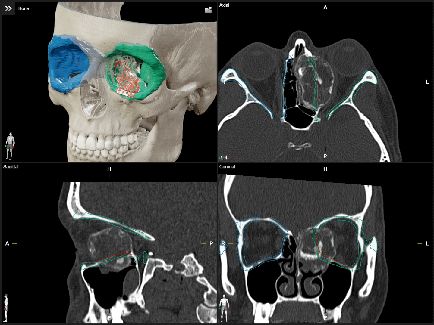
Cover your workflow for CMF planning with Brainlab Elements software. Achieve straightforward patient data alignment, image fusion, object outlining and segmentation of anatomical structures that can be used as a template for affected regions. STL import and export allow you to easily export patient-specific structures for the production of implants or rapid prototyping models and incorporate the final STL file into the plan.

Screenshot: CMF planning with Brainlab Elements software

Mixed Reality Viewing

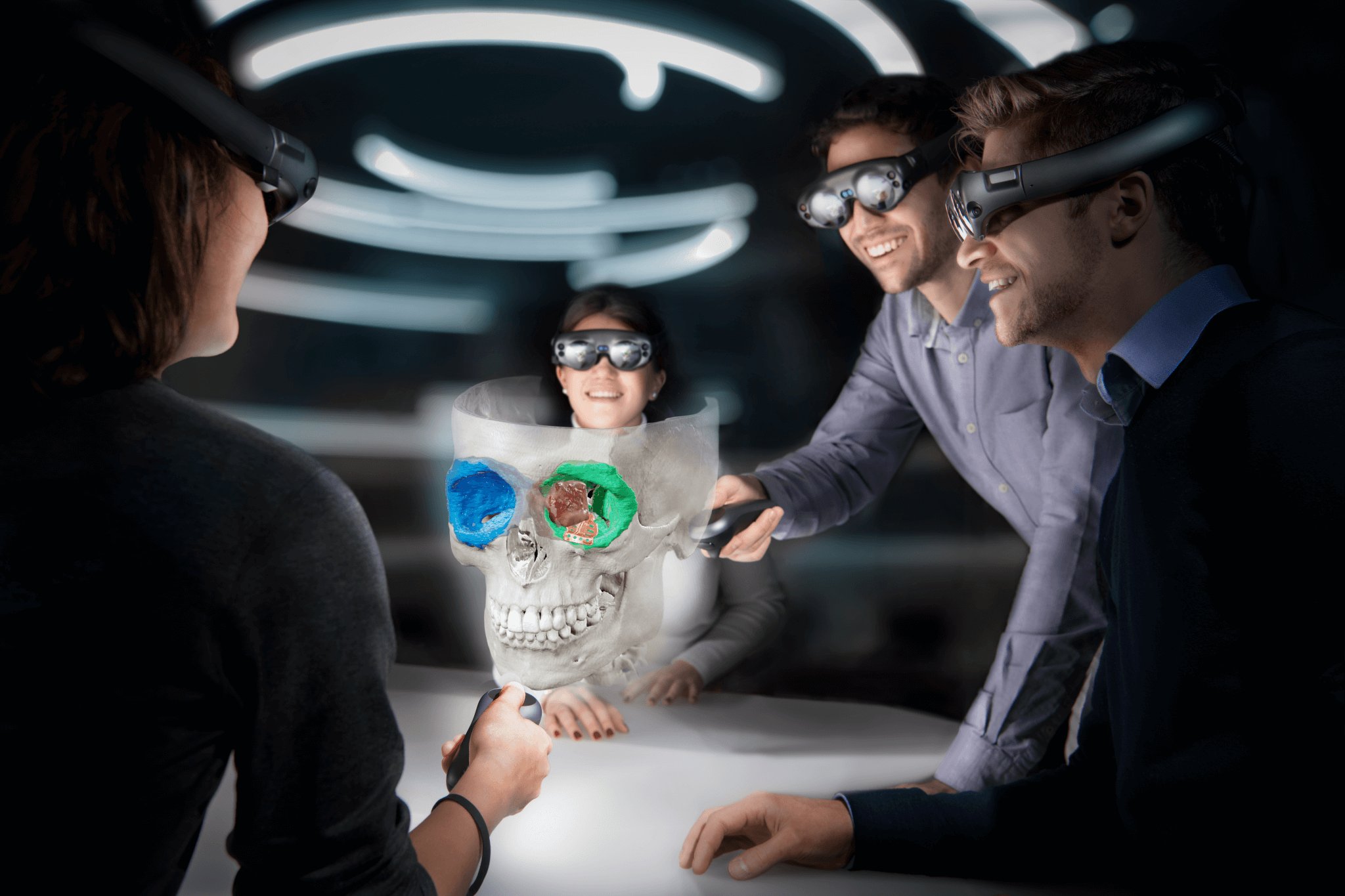
Visualize your reconstruction plan with the Mixed Reality Viewer and dive into the patient’s anatomy in hyper-realistic 3D. Gain deeper insights into case-specific anatomy and the spatial relationships between bone, implant, critical structures and tumor. Virtually size and place your 3D model and discuss the surgical approach together with colleagues or patients.

A group of people wearing the Mixed Reality Viewer gather around a table, examining a cranial image

Surgical Navigation

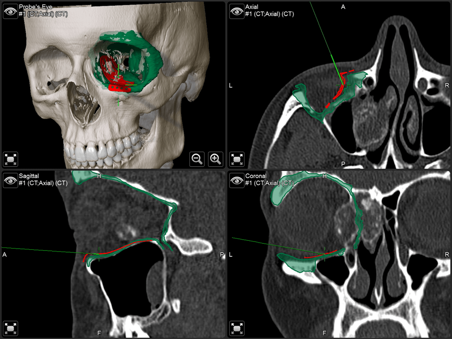
Make the most of your planned data by transferring it into the operating room. With navigation1, final positions of implants, meshes and bone fragments can all be verified. Surgical results can be documented using the structure acquisition functionality, which allows for the intraoperative visualization of the implant surface, including biocompatible materials.

Screenshot: Surgical Navigation with Elements

Intraoperative Imaging

Combine surgical navigation with Loop-X Mobile Imaging Robot or third party imaging systems to instantly verify your progress in the operating room. Acquired intraoperative scans can be fused automatically with the preoperative plan enabling fast and precise quality control for every case.

Teaser Image: Loop-X Mobile Imaging Robot

Let your surgical work flow

How Brainlab technologies support craniomaxillofacial workflows2

Intraoperative imaging can prevent revision surgeries through instant progress verification in the O.R. It enables me to fuse the acquired dataset with my preoperative plan to see if I reached the desired outcome.

Prof. Julio Acero, MD, DMD, PhD, Ramón y Cajal University and Puerta de Hierro University Hospitals, Madrid, Spain
Prof. Julio Acero, MD, DMD, PhD, Ramón y Cajal University and Puerta de Hierro University Hospitals Madrid, Spain

Elements planning software allows me to preoperatively decide on the best possible treatment for my patient. It supports me with patient-specific automatic segmentation of all relevant structures and reconstruction with implants to plan the optimal treatment outcome.

Michael P. Grant MD, PhD, FACS, Craniofacial Surgeon, Baltimore, USA
Michael P. Grant, MD, PhD, FACS, Craniofacial Surgeon, Baltimore, USA

Craniomaxillofacial planning software improves my daily work especially in complex mid-facial reconstructions and tumor treatments. Additionally, the transfer of the planned data to intraoperative systems like imaging or navigation platforms increases my clinical outcome as I can perform a live intraoperative check of my preoperative plan.

Professor Alexander Schramm, MD, DDS, University and Military Hospital Ulm, Germany

Preoperative planning with the Mixed Reality Viewer will give us the ultimate view into virtual reconstruction from standard orbital implants to complex mid-facial fractures. A final check of the implant and all close-by structures will help surgeons to ensure the best possible patient treatment.

Majeed Rana, MD, DMD, Düsseldorf University Hospital, Germany

I see the Mixed Reality Viewer as a major innovation in patient education for head and neck oncology, craniomaxillofacial reconstructive surgery and traumatology.

Prof. Bernd Lethaus, MD, DDS, University of Leipzig Medical Center, Germany

Brainlab navigation enables us to realize preoperative plans. All relevant data is immediately available and can be utilized during surgery. This allows us to perform reconstructive procedures with less radiation for high precision verification of the final implant position.

Prof. Christian Freudlsperger, MD, DDS & Reinald Kühle, MD, DDS, Heidelberg University Hospital, Germany

Intraoperative imaging can prevent revision surgeries through instant progress verification in the O.R. It enables me to fuse the acquired dataset with my preoperative plan to see if I reached the desired outcome.

Prof. Julio Acero, MD, DMD, PhD, Ramón y Cajal University and Puerta de Hierro University Hospitals, Madrid, Spain
Prof. Julio Acero, MD, DMD, PhD, Ramón y Cajal University and Puerta de Hierro University Hospitals Madrid, Spain

Elements planning software allows me to preoperatively decide on the best possible treatment for my patient. It supports me with patient-specific automatic segmentation of all relevant structures and reconstruction with implants to plan the optimal treatment outcome.

Michael P. Grant MD, PhD, FACS, Craniofacial Surgeon, Baltimore, USA
Michael P. Grant, MD, PhD, FACS, Craniofacial Surgeon, Baltimore, USA

Shape the face of your CMF surgeries with digital technologies

Open to Everything

Brainlab has always had an open framework that allows free choice. This open system lets you incorporate the devices and implants you already use into the Brainlab CMF environment including standard and patient-specific models. You’re free to choose the technology that works for you.

Explore more about digital CMF surgery

Brainlab CMF Webinar Series

Join our live CMF webinar series presented by leading clinicians in the field of craniomaxillofacial surgery or discover our collection of on-demand videos to learn more about the latest advances in CMF surgery.
br_brain_xparticlesbaked_v027-fullhd-journal.jpg

Brainlab Journal

Interested to gain more insights about O.R. trends or new industry perspectives? Dig deeper and explore our Brainlab Journal covering topics from artificial intelligence, mixed reality to operating room integration and much more.
  1. Not yet commercially available in several countries (CE mark available). Please contact your sales representative.

  2. The statements of the surgeons reflect only personal opinions and experiences and do not necessarily reflect the opinions of Brainlab or any affiliated institution.