Webinar

Complex Case Discussion Focusing on Navigation and Image Fusion in Sinonasal Tumors

Topics
Intraoperative Imaging
耳鼻喉科
Surgery Navigation
语言

English

共享此页

描述

Brainlab invites you to join a live webinar, “Complex Case Discussion Focusing on Navigation and Image Fusion in Sinonasal Tumors”. Taking place on March 10, 2021 at 4:00 PM CET, this webinar will be presented by Frank Haubner, MD, Senior Physician, Department of Otolaryngology, University Hospital Munich, Germany.


We look forward to meeting you online!

Language | English

In case you can’t join the webinar, it will be recorded and shared afterward.

Participation is free of charge.

The views, information and opinions expressed during this presentation are the speaker’s own and do not necessarily represent those of Brainlab.

演讲人
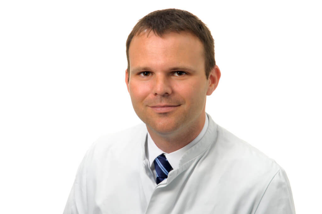
Priv. Doz. Dr. med. Frank Haubner

Senior Physician, Department of Otolaryngology, University Hospital Munich, Germany

主持人

Jana Neider
Jana Neider

Brainlab AG

To watch the webinar, please enter your contact data Please confirm that you would like to register for this webinar

    We offer our webinars free of charge. In return we ask for your permission to receiving promotional emails.

    您可以在 Brainlab 隐私政策中查看关于我们如何处理您的个人数据的信息。

    Are you interested in an easier and faster way to register for upcoming events?
    Create a Brainlab ID account and save time before the next session. Gain access to MyBrainlab Online Services for more related event and webinar recommendations.

    感谢您注册!现在您可以查看录制的数据。

      To join our webinar free of charge, please confirm that we can contact you with related promotional material in the future.

      Information on the processing of your personal data can be found in the Brainlab privacy policy

      Thanks for your registration! You can now view the recording.

      Video Transcript

      Jana: Welcome to our next ENT webinar in our ENT Webinar Series. My name is Anna Nida [SP] and I’m very happy to meet all of you here today. Before I introduce you to our speaker, Dr. Haubner, I would like to explain a few points about this webinar. Today, our webinar focuses on the complex case discussion focusing on navigation and image fusion in sinonasal tumors. A very exciting topic with a very complex structure and a case where you need high precision, so I’m very happy about the interesting lecture coming up. The lecture will last about 45 minutes, followed by a question and answer session of around 15 minutes.

      You can submit any kind of question through the online chat function, and I will address them to Dr. Haubner after he finishes his presentation. This webinar is live, but it will be recorded to be watched again by all registered participants. For further questions, feel free to use the chat function. Now, onto our speaker, Dr. Haubner. Dr. Haubner, thank you for being here today. It’s a pleasure for us to see you and to hear you today and having you speaking about that exciting topic.

      Dr. Haubner is at the University Hospital in Munich. Since 2017, he’s the Senior Physician as the head of the section for rhinology. He also leads several courses and also especially the paranasal sinus course in Munich. He studied at the university in Regensburg, as well as in Cape Town in South Africa. And he also did his PhD at the University of Regensburg.

      Dr. Haubner is very well known for his membership also in several societies. He also is working the German Oto-Rhino-Laryngology Society, as well as Head and Neck, and he was also one of the award winners in 2013 and 2014. Now, I would like to handover to Dr. Haubner. Thank you for your interesting presentation. And, yeah, we are looking very much forward to that.

      Dr. Haubner: Yeah, thank you for the kind introduction. We will share my screen now. It’s my pleasure to be with you today and to discuss with you some advanced cases of tumors within the paranasal sinuses. First, we start with a disclaimer. The views, information, and opinions expressed within this presentation are my own, and so they do not necessarily represent those of Brainlab. Furthermore, I would like to disclose that I have received speaker honorary and travel grants within the last years by those companies. With that, we start with the agenda for today.

      I would like to give you an overview about different entities of tumors within the paranasal sinuses, there are benign lesions as well as malignancies. I will show you the staging system for malignant tumors. We will talk about the preoperative considerations, the different surgical approaches which have to be individualized for each patient. And we will have some time to talk about the role of imaging and navigation in tumor surgery. Then, we will go into detail and discuss some surgical cases and finally, we have some time to answer your questions.

      Let’s start with an overview about the most common benign lesions you can find within the paranasal sinuses. Today, we will focus on two special entities, the inverted papilloma, and the juvenile angiofibroma, because from my point of view, they are the most challenging ones. Furthermore, as you all know, there are also osteomas, different fungal diseases, pyoceles, mucoceles, and of course, polyps within the paranasal sinuses.

      As far as malignancies concern, there are different tumors and the most common are the squamous cell carcinomas. You can also find adenocarcinomas, especially woodworkers. We will see that later on. And then there are rare tumor entities like melanoma, aesthesioneuroblastoma, the ACCs, and neuroendocrine carcinomas.

      If you talk about malignancies, it is always important to have a staging system. And as you know from other tumor entities, there is a TNM classification system also for malignant tumors in the paranasal sinuses. And let’s go through this classification because it is very straightforward and important to know and to remember.

      A T1 tumor is restricted to one area of the nose or to one paranasal sinus. A T2 tumor has an extension into two areas. T3 means that there is an infiltration of either the medial orbit, the orbital floor, the maxillary sinus, the cribriform plate, or the hard palate. Then there is T4a and the T4b classification. T4a means that the tumor infiltrates one or more structures like the orbit, the skin, the anterior skull base, the pterygoideus process, the sphenoid, or the frontal sinus.

      T4b means that there is a tumor infiltration of, for example, the orbital apex, the dura, the brain, the middle cranial fossa, cranial nerves, [inaudible 00:06:46] V2, the nasopharynx, or the clivus. Just keep that in mind as we go later on into detail and discuss the cases.

      It is always important that you have a straightforward workflow for your decision-making in the management of tumors within the paranasal sinuses. For us, that means that we start with a clinical examination of the patient. It is very important that this includes nasal endoscopy. We need imaging and, in tumor cases, I do always recommend CT and MRI scans. It is important to have biopsies and a histopathology examination before you go for surgery in many cases because it depends on the organ staging in case of malignancy. And you will have to exclude or determine locant and distant metastases, and you can only plan your exact treatment if you have a biopsy before.

      Then, in many institutions, so also in our institution, we have the skull base and the tumor board where we can discuss those tumors in an interdisciplinary team with ENT colleagues, colleagues from the neurosurgery department, and especially from the radiotherapy and oncology department to manage and to plan the individual therapy of the patient.

      This board will give a recommendation and this recommendation has to be discussed with the patient or with the family of the patient as far as the further treatment concerns. Then, it can either go into planning of the surgery and go for surgical approaches, or planning or radio or chemotherapy.

      Today, we will like to talk about surgical cases. And if you plan your surgery, you have to think about what kind of approach you would like to use. And many tumors of the paranasal sinuses can be addressed by endoscopic approaches today. But, as you will see later on, also the bold, open approaches can be very helpful. And in my point of view, it is very helpful to combine both approaches, especially in the advanced cases.

      Then, you have to think about, do you need an interdisciplinary approach or an interdisciplinary surgical team, for example, together with a colleague from the department of neurosurgery? And you have to think about the need for navigation or the need for additional imaging in advance. It is important to make sure that all of your imaging you would like to have is available for the day of surgery.

      Now, let’s have a look to the recommendations to use navigation. And there is a quite clear recommendation from the American Academy of Otolaryngology Head and Neck Surgery, that navigation is very helpful and useful in tumor surgery. As you know, also in other cases like revision surgery or situations with an anatomical variations, severe recurrence of polyposis, and involvement of disease to the skull base, orbit, or close relationship to the carotid artery and optic nerve, in all of those situations, is navigation helpful, but there is a strong recommendation to use it in tumor cases.

      Here at my institution, we use this kind of optic navigation system in our OR together with those screens. And from my point of view, it’s very helpful to have this kind of setting in the OR because in interdisciplinary cases, you can discuss the imaging before starting the surgery in the team, and you can discuss special landmarks, and maybe focus specific points for the later resection.

      In many of our cases, we use this headband for calibration of the navigation system. If you work together with the neurosurgeons, you can also use this optical navigation system together with the Mayfield [SP] clamp. Then, let’s come to the question, do we need or when do we need image fusion? And from my point of view, image fusion is very helpful in cases where you cannot really see the extension of the tumor in the CT scan. You see that the additional information of the MRI with the extension of the tumor to the brainstem is only visible in the MRI. And if you go for surgery or biopsy of this tumor, it is helpful to have both information available, and then for me, this tool of image fusion is very helpful and essential to do the case.

      We do a lot of chordoma surgery at our institution. And also, in those clivus chordomas, it is very helpful to use image fusion, because you only see this close relationship to the internal carotid artery in the MRI picture. And you also need the information from the exact bony structures within the sphenoid sinus and the clivus from the CT. So you need both information and this is possible with the tool of image fusion.

      Again, another case of a clivus chordoma with a close relationship to the basilar artery. And if you go for surgery, it is important for you to have the bony landmarks here, the lateral sidewall of the sphenoid sinus, and here, the anterior border of the clivus. But you would also like to see where the soft tissues and the blood vessels. And so again, image fusion of MRI and CT scans is very helpful.

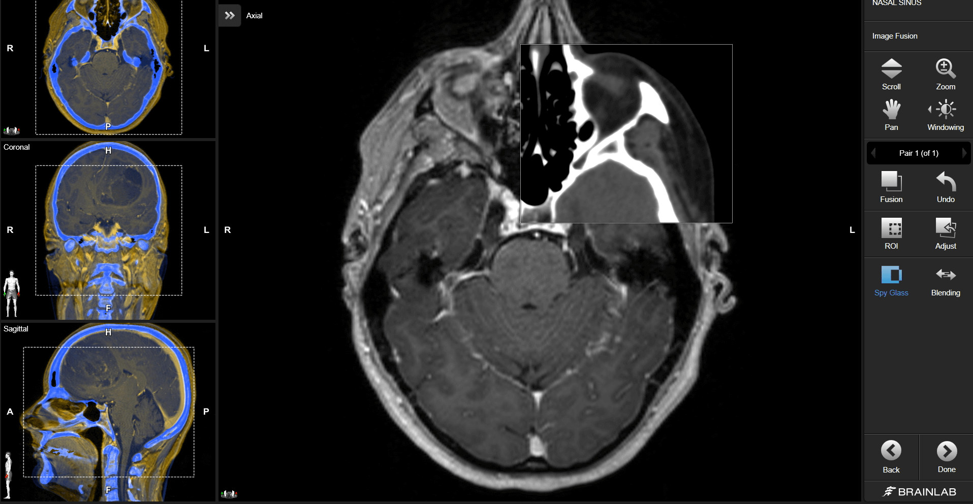
      Now, how to perform image fusion? There are different and multiple possibilities, and yeah, there are software tools, and you can use this software for the fusion mode, and then you can see here, in this so-called spy window of the fusion of the two pairs if this was exact, and you can adapt it to your individual case and situation with this multi-planal [SP] approach. If you are interested in how to do that, you can have a look to the websites and have a look to the instruction of video sequences.

      Now, let’s come to the surgical cases. And as I told you before, I would like to share with you endoscopic approaches and approaches which are combined. That means, for me, an open end, and endoscopically approaches to tumors within the paranasal sinuses. Let’s start with case one. This is a case of a 51-year-old male. He reports about troubled breathing through his nose. He had a previous treatment due to nasal polyps and this is the endoscopic view to his left olfactory cleft. And here, you see that it’s not a polyp, it is a reddish tumor. And biopsy revealed an aesthesioneuroblastoma. Aesthesioneuroblastomas are rare, and very aggressive malignancies of the paranasal sinuses. That is why an additional PET CT scan was performed to exclude local or distant metastasis.

      Before starting the surgery, additional MRIs were performed and that imaging was discussed in our skull base board. For me, it is important to point out that you need the information of the T2 and the T1 sequence of the MRI to clearly see where is tumor issue and what is a cystic component on the tumor, head of the tumor tissue below. And you need both information and you need the information of the additional CT scan to plan and go for surgery.

      The skull base board recommended surgery in this situation, and if you go to surgery of an aesthesioneuroblastoma, you always start with a drill-out procedure and a so-called [inaudible 00:18:04] procedure with a view to both frontal sinuses. Here, the cranial part of the nasal septum is already resected, and then you go on to resecting the tumor tissue along the cribriform plate and the skull base.

      Here, again, image fusion and intraoperative navigation is mandatory, from my point of view, to address also the cystic components of the tumor intracranially. These are now some intraoperative pictures during tumor resection, and it is important to clearly visualize and coagulate the anterior and posterior ethmoid artery and then go for resection of the cribriform plate together with the tumor.

      So, after endoscopic resection skull base, reconstruction was performed by using several layers of DuraGen, a collagen matrix together with TachoSil, a fibrin glue, and additional nasoseptal, a so-called Hadad flap was performed to close up the skull base to the nasal cavity. Final tumor staging showed again an aesthesioneuroblastoma advanced disease Kadish C without any local distant metastasis. And it’s always in those advanced situations with infiltration of skull base to our brain, there’s a recommendation for adjuvant radiotherapy.

      Then, this is his three-month follow-up. You still see this reactive swelling of mucosa within the maxillary sinus. This is due to the radiotherapy and settles down over time. His one-year follow-up, with a nicely healed skull base reconstruction and the recovery of tissue swelling within the paranasal sinuses.

      Let’s come to case two, an 18-year-old young female. She tells you about a blocked nose and if you have a look to her nasal cavity, you see this huge mass at the posterior part of her nasal septum blocking the entire nasopharynx. And this is a rare tumor, this is a neuroendocrine carcinoma. And you have to know that, in this situation, surgery is not the most important thing. It is not so difficult to remove the tumor from the posterior end of the nasal septum because there was no infiltration to the other structures. But the most important treatment of this entity is chemotherapy.

      Let’s come to another surgical case. As I said before, you always have to think, often, adenocarcinoma if you are confronted with woodworkers and tumors within the paranasal sinuses. So this is a 52-year-old male woodworker. And he reports, again, about troubled breathing, and he suffers from this huge mass within his nasal cavity and paranasal sinuses with infiltration of the skull base and the medial and inferior orbital wall.

      Staging was completed without any metastasis, and again, there was a board recommendation for trans-nasal resection. Here, again, it is very important that you have both information available, the MRI and the CT scan, because only in combination of both, you see again, there is a trapped fluid or a cystic component of the tumor next to the tumor tissue, that you cannot see in the CT scan. But is very helpful to have also a CT scan to see this skull base infiltration, but also a small bony landmark along the orbital walls.

      So let’s have a look to the intraoperative situation. This is a typical picture of an adenocarcinoma and this is already after the tumor resection. You see clear fluid coming out of the brain. This is a CSF leak due to tumor infiltration of the dura. And so, we had to reconstruct the dura in this area using DuraGen as underlay and to complete the closure to use some pieces of collagen matrix with fibrin glue to make the closure water-tight.

      Then, this is his post-op scan, and you see this reconstructed area of the skull base without any residual tumor tissue. But due to the extent, we have a very advanced disease with a T4b due to dura infiltration [inaudible 00:24:19]. So there is, again, a clear recommendation for post-op radiotherapy.

      Then, as I said before, the most common entity of the malignancy within the paranasal sinuses is the squamous cell carcinoma. And again, a very advanced case with tumor infiltration, not only of the nose and to the paranasal sinuses but also of the skin envelope of the nose and forehead. You can also see that there is an infiltration to both of the orbits and an infiltration to the skull base. So a very difficult situation where you can and have to discuss multiple strategies, conservative strategies like primary radiochemotherapy, and also of course, surgical approaches where you have to think about and discuss the option of an exenteration orbital on both sides.

      Here, the recommendation was go to surgery. And we planned the surgery and discussed all of the options with the patient and his family. It was important also to point out that we have to resect parts of the skin in this area where the infiltration was. And here, all ready for reconstruction, a pericranial flap was marked. Then, we started with an open resection. That means we took out the tumor tissue together with the infiltrated skin from the nasal cavity, and then we switched over to the endoscopic approach for the further resection along the skull base and the intracranial tumor parts.

      Also, reconstruction is very helpful not to do only in the open approach, but also with a good vision here with a microscopic view where the DuraGen matrix is already covered on the brain tissue. Here, you can see the brain tissue of the frontal lobe, and then it is closed in one layer. And due to the huge defect, we decided to go for additional reconstruction using a titanium mesh fixed here on the anterior border of the frontal sinus wall, and go all the way back behind to the remnants of the sphenoid plane.

      This is another intraop and then the area is covered with a pericranial flap. The final histopathology reports about, in that case, sinonasal undifferentiated carcinoma. A very aggressive one, with a T4b [inaudible 00:27:59] due to brain and dura infiltration. And then there was a clear recommendation for radiochemotherapy in this situation.

      Just to give you some more literature on this topic of sinonasal malignancies. There is a nice article in “Cancer” from 2017 and also a review in “Rhinology” published. And the interesting thing is that there is currently no real standard therapy. You always have to discuss the individual situation together with the patient and his family. But from my point of view, there is quite common sense, especially for the sinonasal carcinomas to go for cross-tumor resection and then post-op radiotherapy.

      Then, let’s switch to other benign tumors in the paranasal sinuses and one very challenging and interesting one for me are the juvenile angiofibromas. And always, if you have young males suffering from recurrent nosebleeds with masses in their nose or paranasal sinuses, you have to think about this rare entity of juvenile angiofibromas. These tumors are not malignant, but they show aggressive behavior due to infiltration of the surrounding tissue, and they have a very high vascularization and that is why they are so difficult to treat.

      Here, a tumor with a more posterior localization with this enlargement in the retromaxillary space, and destruction of the lateral wall of the maxillary sinus.

      This is the MRI of the patient. Again, you can see this lateral extend of the tumor, and also the extension up to the nasopharynx and the skull base. Due to the high vascularization, there is a strong recommendation for pre-op embolization of those tumors, and that was all…

      [00:30:53]

      [silence]

      [00:31:14]

      Dr. Haubner: We used some of the surgical steps. You start with a C-shaped incision. This is a left-side C-shaped incision along the inferior turbinate. And then you go subperiosteal to the bone of the turbinate and you will have then the possibility to take out the bones from the inferior turbinate as well as parts of the anterior wall of the maxillary sinus.

      Then, you can push all the soft tissue together with the nasolacrimal duct here to the midline, and get access to the left maxillary sinus. You have now an excellent view to the posterior wall, the lateral wall, as well as the orbital floor. Then you see the tumor pushes here into the maxillary sinus. There is still the pulsation and you have now an excellent visualization due to this prelacrimal access where you can see [inaudible 00:32:41] you can have a look to the coiled maxillary artery. And you can take out the entire tumor endoscopically through this access.

      From my point of view, this prelacrimal approach is very elegant and very helpful because it has a very less morbidity. You can just bring back the inferior turbinate to the lateral nasal wall, put some stitches, and you do not have a very extended wound area.

      But there are also other cases of JNAs where, for example, like in this situation, the whole nasal cavity is filled up with tumor tissue. And for me, this is my opinion, it is hard to plan an endoscopic approach if there is no room for your endoscope at the beginning of the surgery. Here, you have a huge extension far lateral, as well as an extension along the skull base with close contact to the vessels and also close contact of the tumor to the cavernous sinus.

      You can see that this is a benign lesion, but it shows aggressive and destructive behavior, if you have a look to the CT and the destruction of the bony structures is visible in the CT scans. As discussed before, we always recommend pre-op embolization to reduce the blood flow in the tumor tissue. In this case, the embolization was performed with Onyx. And as I said, it is hard to start with an endoscopic approach if you do not have room for your endoscope. That is why I decided to go for a midfacial degloving in this situation. And that means that you can develop the soft tissue and skin envelope of the face, and you have a good visualization to both maxillary sinus walls as well as the tumor in the nasal cavity.

      I always use pre-plating. That means that before I remove the bony wall of the maxillary sinus, I plan my reconstruction with those titanium plates, and then I take out the plates and use them later on for reconstruction. Then, the anterior wall of the maxillary sinus was removed and you have an excellent view to the tumor and it is possible to develop and resect the lateral mound of the tumor very bluntly and under good vision.

      After mobilizing this part of the tumor, we switched over to an endoscopic approach, because I think if you go to tumor resection along the skull base and especially in close neighborhood of the huge vessels like the internal carotid artery, you have a better vision of the depth surgical field, if you use your endoscope.

      And here, again, from my point of view, image fusion is very helpful, because you see the bony structures here in the sphenoid sinus, and you have a very excellent orientation during the resection of the tumor. Because orientation is very important, the surgical field can be very bloody. And so for me, navigation including image fusion is helpful or even mandatory.

      Then, you have to identify the tumor origin. And as you probably know, the tumor origin of the JNAs can be the pterygoid route and that was also the case in this situation where you see small remnants or nests of tumor tissue along the pterygoid route and you have to drill them off and sharply dissect to avoid tumor recurrence from this area. And again, to identify the pterygoid route, it is very helpful to use the navigation system.

      This is then the intraop situation already with tumor resection performed from the pterygoid from sphenoid sinus is opened up and you see that there is clear surgical field along the skull base. The sphenoid sinus, the posterior end of your nasal septum was here, partially resected, and the tumor was taken out of the nasopharynx. You see both two ostiums on each side.

      Then the tumor was sent to histopathology examination. And as I said before, those JNAs are not malignant tumors, but you have to consider that recurrences can occur in those advanced stages, and we always recommend to perform MRI scans after six months within the first two years after tumor resection. Then, the reconstruction was performed using titanium plates and the bone which was taken before.

      Then, let’s come to another benign lesion, which can be very challenging, these are the inverted papillomas. Inverted papillomas, they belong to the benign tumors or the paranasal sinuses, but you have to treat them like malignancies. That means you need complete resection of the tumor, otherwise, there will be a recurrence of the tumor end. Furthermore, you have the risk of development of malignancies within the inverted papillomas in about 5% to 10% of the cases. So complete surgical resection is very important. And as you can imagine, if you have a situation like that with infiltration of the lateral wall of the maxillary sinus and infiltration of the palate, this is challenging and you can think about using an open approach, maybe together with the colleagues from the maxillary facial surgery department. Or, you can also think about using an endoscopic approach. And in this situation, we decided to use an endoscopic approach. Again, supported by a navigation system and image fusion to get excellent visualization and support during the tumor resection.

      You start by taking out parts of the tumor from the nasal cavity to get access into the nose for your endoscopic. And then, a so-called hemimaxillectomy was performed here with resection of the nasolacrimal duct. You see the tears coming out of the duct, and then you go for your resection along the orbital floor, all the way lateral until you meet here, the buccal fat. And then, the tumor was taken out entirely from this lateral dimension and the bony wall of the maxillary sinus was drilled off with a diamond burr.

      So, let’s come to the last case for today. A very rare case I would like with you. This patient suffering from an arachnoid cyst. These are benign lesions filled with CSF, but they grow over time and can deform even the bone, here, of the anterior wall of the frontal sinus. And they lead to headache because they have a lot of pressure to the brain at the frontal lobe, and that was the situation in this gentleman who presented with headache due to this arachnoid cyst.

      In a situation like that, you can use a bicranial approach, that would be probably the standard approach in many centers, a bicoronal incision, and then you have an excellent view to the frontal lobe, and you can take out the tumor tissue, have good access for reconstruction. But, you know, it is a benign lesion and we always would like to avoid morbidity. That is why I decided to use this transorbital approach, and I would like to share the surgical steps with you.

      You can use the supratarsal incision, which is very, very elegant because you do not have a lot of scarring in this area. It is similar to blepharoplasty incision where you can really hide your incision line. Then, you go for subperiosteal preparation and dissection of the orbital rim. And as you can see here, I used, again, this pre-op plating with holes for the screws. And then we used this Piezo [SP] instrument to take out a piece of bone of your orbital roof and then you can take out the bone and have this view. Yeah. And yeah, it is a small window, but it is enough to introduce your endoscopic instrument. This is a very minimally invasive approach for access of the anterior skull base.

      Then, this is the endoscopical view to this patient. You introduce your endoscope to the opening, you still see some remnants of this arachnoid cyst. You see the anterior wall of the frontal sinus. And you see the pulsation of the brain. And you can also use this access for reconstruction. And this was also performed. In this case, we used several layers of collagen matrix together with fibrin glue to cover the skull base and to reconstruct the skull base to prevent further CSF leaking.

      And as you see, you can go really far up into this frontal sinus and you can reach all the areas, lateral, medial to close the defect.

      Then, reconstruction of the anterior wall is also important. As I said before, you can use your plates with some screws, then their fixation is performed. And we always recommended to do a CT scan after reconstruction to show if there is any signs of leakage or free air which can occur. So it’s important to make sure that your reconstruction was successful.

      So that was my last case for today, and let’s come to the conclusion. I think imaging and navigation are essential parts in skull base tumor surgery. An interdisciplinary team is mandatory. And it’s very helpful to have a neurosurgeon next to you. An endoscopic approach can be combined, in my opinion, with open approaches to itemize the surgical field. And imaging including navigation and image fusion are key for safe tumor resection, especially in these advanced cases.

      So, with that, I thank you for your attention. And I’m very happy to answer your questions now.

      Jana: Thank you, Dr. Haubner. That was a very interesting talk, very impressive, interesting images, very key. We already received a lot of questions. And I would like to remind everyone in the webinar, that you can start asking questions. I will still address them to Dr. Haubner. But first, actually, I have a question to you, Dr. Haubner. And the question is actually, how do you utilize navigation during these cases? Are you using navigation to find your right approach or also for the resection control?

      Dr. Haubner: Yeah, actually both. As I showed in the beginning, there are always dangerous structures and we would like to avoid injury, for example, to the internal carotid artery. So it is very important to use navigation for the important landmarks during the resection. And as I also showed in the case of this huge juvenile angiofibroma, it is helpful to show that you have reached the border of resection, and there is no more tumor tissue in the patient.

      Jana: Thank you. I think that perfectly leads to the next question from of the attendees. And the question is, what kind of factors in navigation platform would you describe as the most vital in cases such as the cases that you presented? So where do you see the most, big benefits?

      Dr. Haubner: From my point of view, yeah, you know, there are different and multiple platforms for a lot of companies. From my point of view, there is a benefit to start with this fusion mode of MRI and CT scan at the beginning. It is very important that you have an exact match of both pictures. And I have shown to you this instructional video sequence, and it’s not always perfect on the first try. But you have to work on this very precise fusion and then you have the possibility to accept these pairs of images, the fused images. And then, you start with your surgical case.

      So do that first in the platform, and then start the case. And yeah, if you have problems during the case, you can repeat, of course. But from my point of view, it’s good to start with the fusion first and go on with the surgery later on.

      Jana: One question that we also received is, if you are actually just using navigation in these very specific difficult cases or do you use it regularly in other cases?

      Dr. Haubner: Yeah, as you know, navigation in our institution is available for almost every case. So we use it very often. And I think if you use it often, it’s, for you, easier, because if you have a special case, a tumor case, or an advanced case with special anatomy, and you use it for the first time, it’s always difficult to handle. So my recommendation is to use it on a regular basis, then you are familiar with the tools and you know how to perform and precise calibration.

      Jana: Thank you for the answer. The next question is… Thank you for the wonderful presentation. And is there a difference when you using electromagnetic or optical navigation? What is your preference and what are you using in your hospital?

      Dr. Haubner: Yeah, as I showed you, we use the optics system. From my point of view, I have very good experience with the optical system. Sometimes in the advanced cases, if you go very deeply into the skull base, for example, during clivus chordoma resection, it can be helpful to use the Mayfield clamp because my impression is that the calibration is a little bit more precise than compared to the headband. And I know different systems using the electromagnetic ones, which have other advantages because you do not have the problem with this line of sight effects, but yeah, there are some data and probably Miss Nida knows that better than me, that the optic systems are a little bit better as far as precision concerns. But there was a strong development during the last five years, so also the electromagnetic systems are excellent now. Would you like to comment on this?

      Jana: Yeah, I think… Yes, thank you, Dr. Haubner. So I think there’s not a real big difference anymore between electromagnetic or optic navigation systems. Also from an accuracy point of view, at least for the Brainlab systems, I can speak, it’s the same accuracy that you can achieve with those systems. They both have different advantages. So with the optical system, of course, you don’t have any kind of metal interference, which is nice if you want to especially use a big microscope or you wanna bring in an intraoperative scanning device, for example. But with electromagnetic, of course, you don’t have any line of sight issues. So always, everything is usable. So especially in forehand surgery, this is something very easy to handle and especially if you do a lot of endoscopic approaches, electromagnetic navigation is really key and very helpful in these cases.

      That brings me also to the next question from one of the attendees. And he was basically asking, are you using also intraoperative imaging, because you mostly showed you preoperatively do the image fusion? But what do you think about intraoperative image fusion…intraoperative imaging? Are you using it? And if yes or no, why? What would be the benefit?

      Dr. Haubner: Yeah. You know, it’s nice to have, yeah. And at my institution, we have the possibility in one OR to use intraoperative imaging, but this is not the standard. But if you go into trouble and you think of having maybe a complication and you need, immediately, feedback, then this intraoperative imaging is extremely helpful. But we do not use it for our tumor cases on the regular basis.

      Jana: Thank you. Yeah, I can also agree to that point. Intraoperative imaging, if you have the option to have that available in your OR, of course, brings a lot of benefit because you can do a final resection control. I mean, the navigation gives you the opportunity to handle the case completely radiation-free, but of course, having a final intraoperative imaging shot before you close the patient is always beneficial [inaudible 00:55:49] post-operatively. Otherwise, if you can do that intraoperatively, it’s of course a big value. The next question is very clinical. So in the recurrence of an angiofibroma which lies on the ACI [SP] [inaudible 00:56:02], would you aim to perform another resection?

      Dr. Haubner: Yeah, that depends. But, yes. Yeah. There are only a few other options to treat them. And you can think about radiotherapy, you can, if there are no clinical problems, no complaints of the patient, you can also think of, yeah, watch and wait. There are some first small cases out there, which are treated with anti-steroids, so a conservative, not a surgical approach. But in many cases, if there is a recurrence and you have to treat it, the re-operation would be the choice.

      Jana: Thank you. The next question that we received is, are you always having CT, and are they available? Or do you also just have sometimes an [inaudible 00:57:16] example in pediatric cases? And then, do you also navigate on [inaudible 00:57:22]?

      Dr. Haubner: Yeah, yeah. An interesting and good question. From my point of view, I would like to have a CT scan before starting the surgery. And so, I always recommend to do CT scan, because the bony landmarks also in children are very important for me to see them and to see if there are any signs of destruction. And I need, for my orientation and to have a safe surgical plan, the CT scan.

      Jana: Thank you. I think the very last question is actually from my end. And I would like to know, so you showed a lot that you do as preoperative planning and you consult the team how to approach these cases. So what would be your number one software tool that you need in your pocket in order to have better planning capabilities?

      Dr. Haubner: Yeah, you know, there are software tools also from your company, and it is nice to discuss the cases and to draw, for example, the lines of the plan to more resection in the CT or in the MRI before starting the surgery. And yeah, that is very helpful, and to think about the team together, for example, with the neurosurgeon, where we will do our resection lines. And it’s good to plan that before. And then, if you have these drawing tools in the software, this is very helpful to plan.

      Jana: Thank you. I think that’s really helpful for us, and I’m sure we’re gonna have another session on what planning capabilities Brainlab also offers for ENT surgeons. With that very last question, I would also like to close our webinar. Thank you, Dr. Haubner, again, for that wonderful presentation. We received a lot of positive feedback that I will also transfer to you. For all of the attendees, we have recurring webinars. So just always feel free to visit our Brainlab.com/webinar webpage where you can see what kind of webinars are upcoming next. We would also send out an email to you in the days for the next ENT webinar. So there’s a few more coming up, and some also really exciting ones that we wanted to advertise to you in a special way this time. So very much looking forward to that. And if you want more information, just send us an email to [email protected] or follow us on our social media.

      Dr. Haubner, thank you again for that wonderful presentation.

      Dr. Haubner: Thank you.

      Jana: Thank you. Thank you, all of the participants for joining today. It was a wonderful time and thanks to everyone. Stay happy and safe. Goodbye. Thank you.

      Related Webinars

      02Blurred_lights_full
      Image Guided SurgeryCranial Surgery+ 3
      Ultrasound Navigation: Confidence in Tumor Resections

      Brainlab invites you to a live webinar, “Ultrasound Navigation: Confidence in Tumor Resections”, at 4 pm CEST on August 3. This session is hosted by Dr. Aliasgar Moiyadi, MD, at the Tata Memorial Centre, Mumbai for Treatment Research & Education …

      bainlab-loop-x
      Intraoperative Imaging颅颌面+ 1
      Expanding my Craniomaxillofacial intraoperative imaging experience with Loop-X, a mobile imaging robot

      Brainlab invites you to join our live webinar, “Expanding my Craniomaxillofacial intraoperative imaging experience with Loop-X, a mobile imaging robot”, on March 16, 2021 at 4:00 PM CET presented by Simon Enzinger, MD, DMD, Managing Senior Physician at the University …

      Simon Enzinger
      webinar-cmf-9
      Intraoperative Imaging颅颌面+ 2
      Benefits of navigation and intraoperative imaging in Cranio-Maxillofacial Surgery – a clinical case review

      Brainlab invites you to join our live webinar, “Benefits of navigation and intraoperative imaging in Cranio-Maxillofacial Surgery – a clinical case review”, on February 9, 2021 at 4:00 PM CET presented by Prof. Julio Acero, MD, DMD, PhD, President of the …

      Prof. Julio Acero

      See more upcoming webinars

      Register now