AANS 2025

Brainlab at AANS — The American Association of Neurological Surgeons

-
Boston Convention & Exhibition Center
Boston, MA, USA

About AANS 2025

Join us in the historic city of Boston from April 25-28, 2025, for the 2025 American Association of Neurological Surgeons Annual Scientific Meeting.
This year’s theme, Power of One, Impact of Many, celebrates the individual contributions that collectively advance the field of neurosurgery. 

Visit us at booth #818!

Detailed Product Information

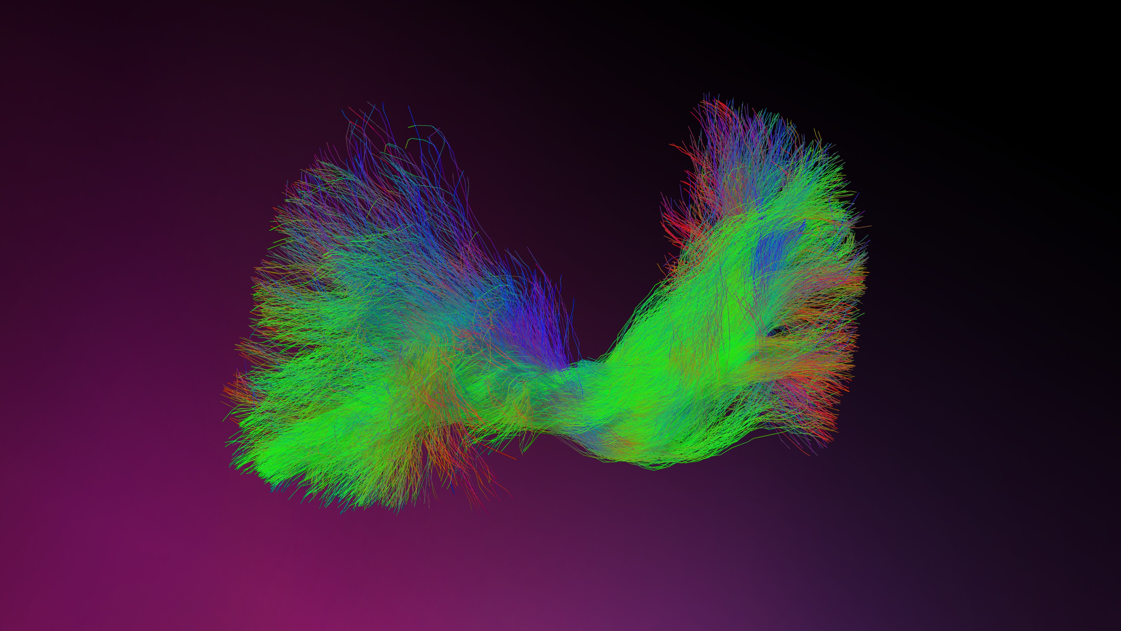
Elements Fibertracking—Matter that matters 

Make informed decisions with advanced tracking1: Take full advantage of research-grade probabilistic tractography in your clinical practice to identify multiple fiber directions like crossing and fanning fibers, as well as fibers that may be present in edema. 

Use tracking templates to expedite your planning: Choose from a selection of tracking templates, (e.g., IFOF1, CST) that enable efficient, standardized and reproducible tracking of important white matter tracts in the regions of interest. 


1
Not yet commercially available in several countries.
 

fbt3-0_keyvisual02_4k-2.jpg

Elements AI Tumor Segmentation1 

Receive a semi-automatic segmentation proposal of diagnosed cranial tumors and segment one or multiple primary brain tumors based on contrast-enhanced T1-weighted MR images powered by the Anatomical Patient Model. 

Detailed quantitative analysis of segmented tumors, including volume, shape and growth rate metrics, provided by Elements SmartBrush. 


1
Not yet commercially available in several countries.
 

240314_elements_surgery_1920x1080px.png

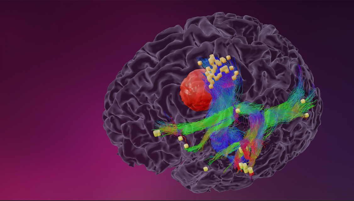
Motor and language mapping with Nexstim nTMS—Preserving what is essential 

Increase resection rates: Preoperative nTMS analysis, combined with nTMS-based fiber tracking, improves the delineation of safe resection limits, leading to increased gross total rection rates and the potential for extended progression-free survival.1,2

Improve clinical outcomes: Using nTMS mapping results as a region of interest facilitates function-based fiber tracking and may improve motor outcomes in tumor resections.1,2

Discover the power of Nexstim nTMS combined with Brainlab Elements for Cranial Planning! Check out our hands-on demo to see how they work together to improve outcomes and find out why nTMS is an essential technique in neurosurgery. 


1
Vucic S. et al., Clinical diagnostic utility of transcranial magnetic stimulation in neurological disorders. Updated report of an IFCN committee. Clin Neurophysiol. 2023 Jun:150:131-175
 

2Krieg S. et al., Preoperative motor mapping by navigated transcranial magnetic brain stimulation improves outcome for motor eloquent lesions. Neuro Oncol. first published online February 9, 2014 

nexstim_aans.png

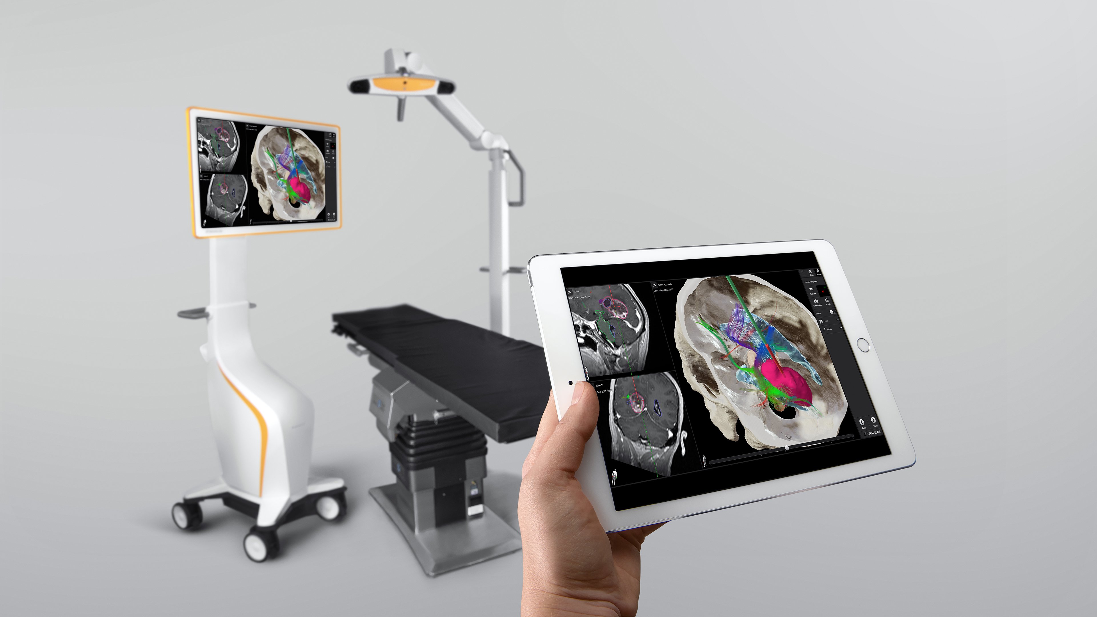
Cranial Navigation—Detailed visualization. Intuitive control. 

When navigating, distraction-free concentration is essential. Detailed visualizations are tailored to show only clinically-relevant data, allowing the surgeon to focus on what’s necessary for their work.  

Informed approach planning requires consolidated clinical data. Cranial Navigation summarizes relevant and detailed anatomical information in one dedicated view, helping surgeons identify the optimal craniotomy before making the incision. 

Origin Remote Control allows for fast and efficient control of the navigation system with a tablet at any time during the procedure. 

ns_img_remote-control-cranial-4k_mar24_rev1.jpg

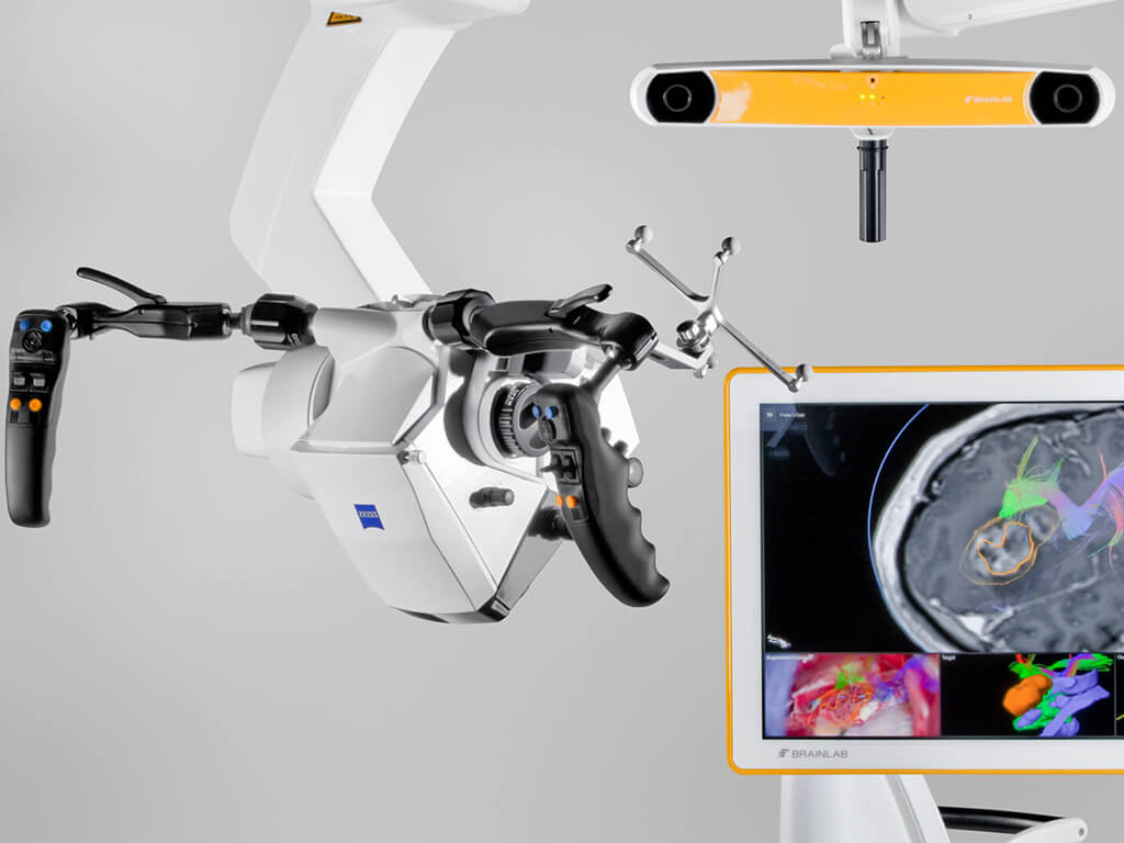
Microscope Navigation with ZEISS KINEVO® 900 S & PENTERO® 800 S 

We’re introducing our brand-new Microscope Navigation integration with ZEISS KINEVO® 900 S1, the latest robotic visualization system, and ZEISS PENTERO® 800 S1, the reimagined advanced visualization system. Both integrations are available for cranial and spinal workflows.

Experience unique augmented reality views, navigation-controlled robotics and the possibility to correct registration errors with our exclusive Navigation Update feature.

1Not yet commercially available. 

Cranial Microscope Integration with Navigation Software

Navigated Disposable Suction 

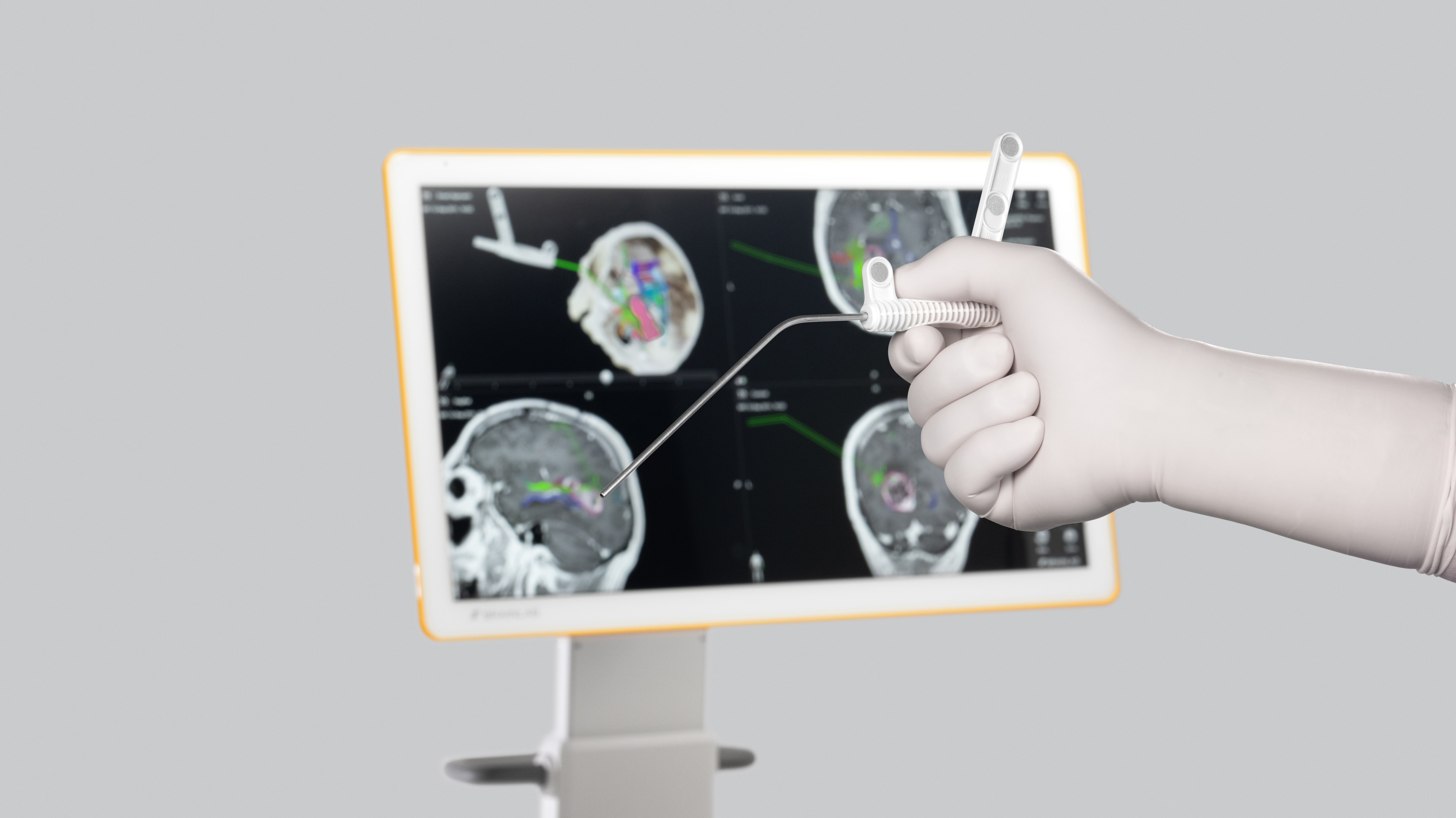
The Disposable Pre-calibrated Suction1 combines the functionality of a classic suction and a navigated pointer into one unique instrument. Its ergonomic, lightweight design supports seamless navigation, especially under the microscope. 

Available with an outer tube diameter of 2.7 mm (Charr 8) and 4.0 mm (Charr 12) and lightweight construction (14 g or 18 g) for comfortable instrument handling. Materials do not contain natural rubber latex, making it suitable for surgeries on patients with latex allergies. 


1
Not yet commercially available in several countries (FDA clearance available).

ns_img_disposable-suction_apr23_rev1.jpg

Ultrasound Navigation—Dynamic layer of confidence 

A whole new dimension. In real time. At any time. We’re delivering innovative, science-backed technology to help surgeons achieve their goal of maximum safe resection, even if brain shift or other navigational inaccuracies occur.  

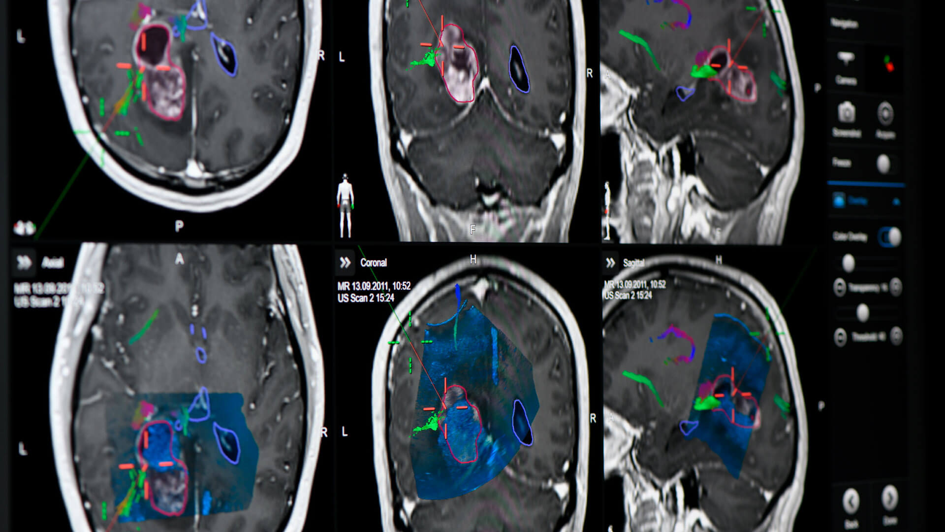
Real-time correlation of intraoperative ultrasound and pre-op MRI: Forget everything you know about intraoperative ultrasound: Gain valuable real-time insights through the right orientation and instantaneous correlation between ultrasound and familiar pre-op MRI images. 

Precision and confidence in navigating cranial resection challenges: Gain the navigational accuracy you need to address the greatest challenges in cranial resection: visualization of and compensation for brain shift, achievement of maximum safe resection and identification of residual tumor. 

Efficient intraoperative imaging without workflow interruption: Take advantage of our cost-effective, fast and straightforward intraoperative imaging technology that will not interrupt your workflow. 

Screenshot of the Ultrasound Navigation: real-time intraoperative imaging for cranial image guidance

Cirq® for LITT—Frameless precision, streamlined workflow 

Now FDA-cleared, Cirq® expands its capabilities to support robotic-assisted alignment and trajectory planning for laser interstitial thermal therapy (LITT1). Designed to work within the Brainlab ecosystem, Cirq® digitizes and integrates the entire process—from planning to execution, from diagnosis to therapy. 

Why it matters? 

  • New compatibility with Monteris NeuroBlate® 

  • Biopsy & LITT in a single streamlined workflow 

  • Real-time tracking & precise guide tube alignment 

  • Robotic-assisted accuracy within an integrated ecosystem 

Experience it firsthand! A special guest will be performing a live demo of our latest technology. Don’t miss this chance to see Cirq® in action! 

 
1Not yet commercially available in the US

Header hero visual for Cirq for Functional Neurosurgery

Cirq® for cranial biopsies—Streamline your biopsy workflow 

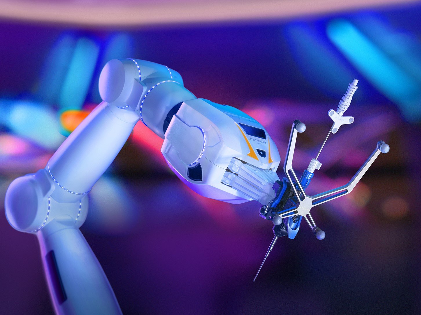
Accurate alignment to the right trajectories: Cirq® boosts alignment accuracy as software visually guides users toward the region of interest. Before the surgery, Elements Trajectory Planning supports in defining the right trajectories. During the procedure, real-time visualization of the biopsy window guides surgeons toward the predefined biopsy target. Other trajectories can also be acquired using a virtual instrument offset. 

Consistent stability and precision: The custom drill guide helps keep incisions minimally invasive while the dedicated biopsy anchor for the skull adds stability to the frameless setup. Cirq® can also track pre-calibrated biopsy needle insertion and biopsy positions can be saved with a simple click of a button. 

Seamless integration with navigation-ready instruments: With navigation-ready instruments, Cirq® is designed to seamlessly integrate into Brainlab planning and cranial navigation solutions as well as new and existing workflows. These features maximize its utility during biopsy workflows and beyond. 

cirq_cranial_hero_0529_colorpurple.jpg

Functional Neurosurgery 

Experience the latest development in functional robotics with the Brainlab Cirq® robotic arm for SEEG. Stop by to learn how this automated robotic platform seamlessly integrates advanced Elements planning and visualization software into the surgical space. The design is compact and surgeon-controlled, offering an efficient solution that utilizes intelligent software features to provide the accuracy you expect for functional neurosurgery while potentially reducing the clinical setup time for your cases. 

Learn how Brainlab Elements offers advanced planning to ensure accuracy and precision at every step of the DBS workflow, from industry-leading Image Fusion, voxel-by-voxel patient Basal Ganglia Segmentation and automated 3D Lead Localization to ensure the highest level of precision in a situation where millimeters matter. 

Our latest developments in functional planning include recent additions to Elements Trajectory Planning. 

Explore unique Brainlab solutions to advance each individual step of your epilepsy surgery procedures, from hypothesis planning to SEEG and surgical intervention. With options for advanced planning and visualization tools, such as Cranial Segmentation, Fibertracking and BOLD MRI Mapping, and robotic platforms for epilepsy surgery and imaging, Brainlab can fit into your workflow at any stage. 

Experience the latest development in functional robotics with the Cirq® robotic arm for SEEG. Stop by to learn how this automated robotic platform seamlessly integrates advanced Elements planning and visualization software into the surgical space with a compact, surgeon-controlled and efficient platform that utilizes intelligent software features—providing the accuracy you expect for functional neurosurgery while potentially reducing the clinical setup time for your cases. 

Image showcasing blue and pink waves and tubes illustrating Brainlab's Functional Neurosurgery Solutions

Robotic Suite 

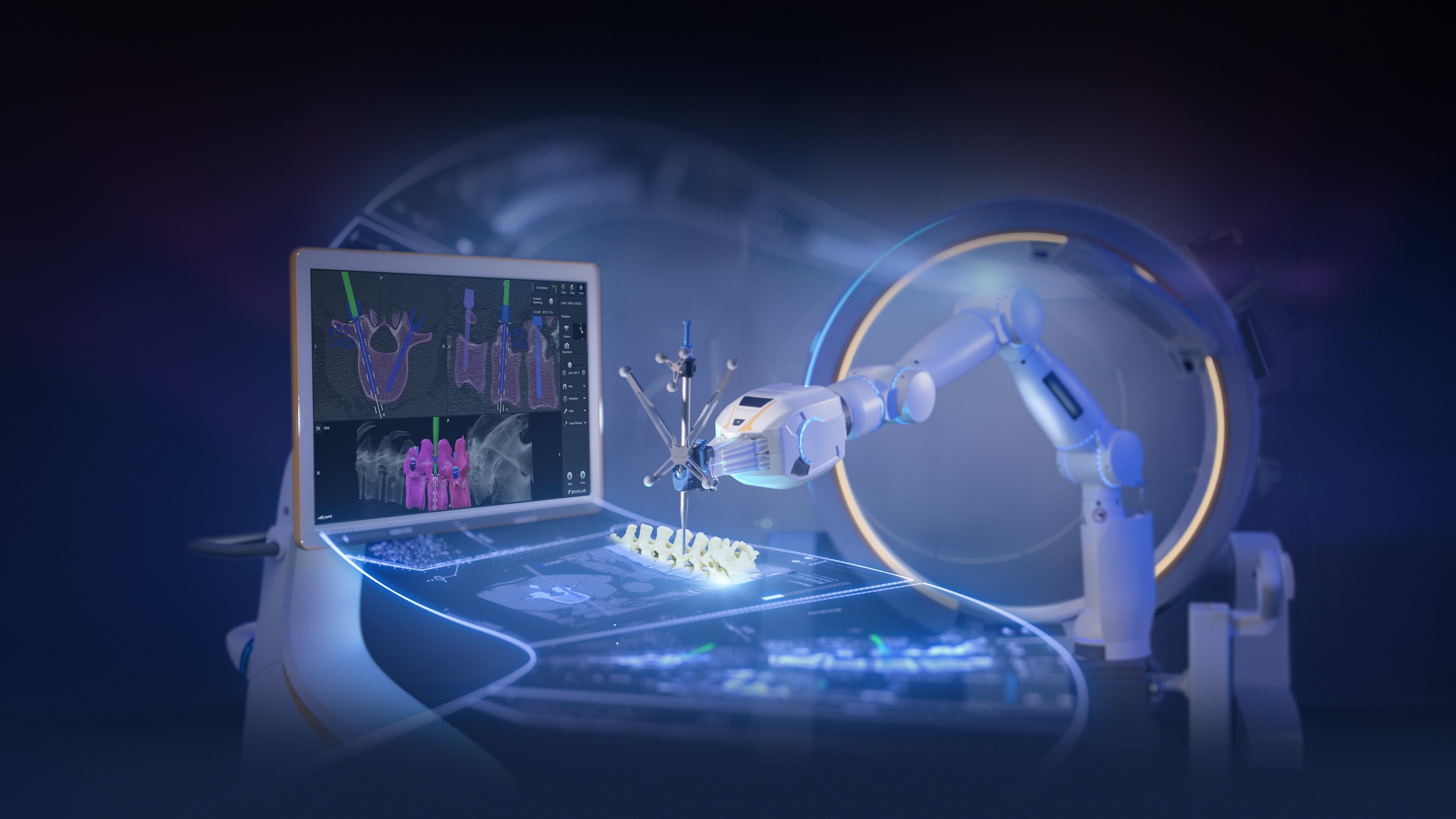
The Robotic Suite creates an ecosystem of accuracy at every step of the surgical workflow by leveraging versatile software to combine three Brainlab products with state-of-the-art modalities: high resolution robotic imaging from Loop-X mobile imaging robot, data processing and precise navigation with Curve Navigation, and automated, stable surgical support with Cirq®, the next- generation robotic arm. 

Image depicting a webinar showcasing Brainlab's robotic suit products

Spine Planning and Navigation 

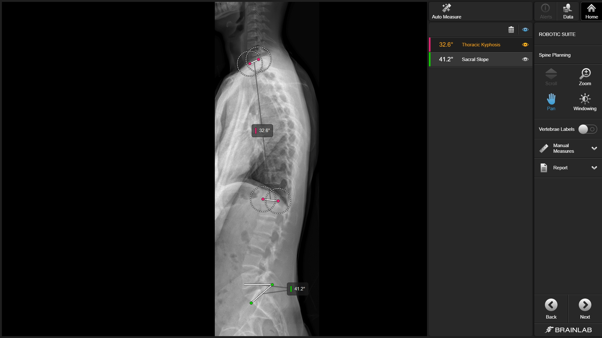
Navigation and robotics have made it clear that screw planning is becoming a necessity and standard practice in spine surgery. With Elements Spine Planning, you can benefit from level detection throughout the complete spine, automated measurements on 2D, and automated screw planning from cervical to sacrum on 3D. The automated functions deliver results and information in seconds, potentially saving time in daily routines and increasing efficiency of entire clinical workflows from planning to logistics to intraoperative data management. Navigation Software Spine & Trauma is the cornerstone of our spine workflow and acts as a central data hub as it seamlessly switches between applications, enabling the versatile surgery setup with Cirq®, Alignment Software Spine and Loop-X mobile imaging robot. Navigation of interbody instruments enables surgeons to navigate every step of their workflow, beyond screw placement. 

Screenshot of the automatic measurements function within Elements for Spine Planning

Mixed Reality Viewer 

Review plans for spine surgery in 3D. The Mixed Reality Viewer1 is a software that works in tandem with Brainlab Elements to produce hyper-realistic 3D patient data. In the context of spine surgery, this software enables medical professionals to review planned surgical procedures as well as explore anatomical structures and planned implant positions. 

1Not yet commercially available in several countries.

2.jpg

Listen to Dr. Thomas Beaumont’s presentation:

“Integrated Multimodal Resection of Eloquent Brain Tumors with Augmented and Mixed Reality” 
 
Speaker: Dr. Thomas Beaumont, MD, PhD 
Time/Date: Saturday, April 26th, 12:15 PM 
Location: Neurohub 
Registration is not required 
Topic: Integrated Multimodal Resection of Eloquent Brain Tumors with Augmented and Mixed Reality 
 
Dr. Beaumont of University of California San Diego will discuss his approaches to eloquent tumors using the latest technology from Brainlab,
including Elements Fibertracking, Augmented and Mixed Reality. Combining the gold-standard in preoperative planning with intraoperative tools
like 5-ALA fluorescence and cortical mapping, Dr. Beaumont shares his multimodal approach to the extent of total resection in diffuse gliomas.

Join us at booth #818 to discover the latest Brainlab innovations

See you at AANS!

Map and Directions

Boston Convention & Exhibition Center
Boston
MA
USA