
Tumor resection demands more than precision. It demands that precision hold as anatomy shifts and conditions evolve. Brainlab connects the brain tumor resection workflow from preoperative planning through intraoperative guidance, supporting informed decisions with the patient at the center.
Resect with confidence
preserve with purpose
Every resection carries a dual commitment—to remove as much of the tumor as possible, and to protect everything that makes the patient who they are. Our integrated workflow supports both, at every stage of the procedure.
Plan with precision
connect every step
Successful tumor resection starts with precise preoperative planning. Our solutions bring together anatomical and functional information, navigation, intraoperative imaging and neuromonitoring so each technology can contribute at the right moment. Together, they carry relevant information from the first plan through the procedure, creating a coherent workflow in which every step matters.
Track progress
guide decisions
As tumor resection progresses, the anatomy and information in view continue to evolve. Our solutions provide intraoperative guidance and updates to help review anatomy, assess resection progress and guide your next decision.
Make every step count
Learn how an integrated cranial workflow can bring planning, navigation, imaging and functional information together for tumor resection. Speak with our team about the needs of your practice.
From plan to reality—
the complete tumor resection workflow
A successful resection begins long before the first incision. It begins with understanding—of the tumor, of the anatomy surrounding it and of the patient whose function depends on every decision made. Our workflow connects each phase of that journey, from the clarity that's built in preoperative planning to the real-time intelligence that guides you through resection.

Understand patient anatomy before you begin
The decisions you make before entering the O.R. shape everything that follows. Brainlab preoperative planning workflow brings together structural and functional imaging to support an informed surgical strategy before the first incision.


Accurate from the start,
adaptable throughout
In tumor resection, navigational accuracy from the first moment is critical—the difference between a confident approach to the tumor margin and one that leaves room for doubt. Brainlab offers registration methods for different O.R. setups, from automatic registration with Loop-X3 and other intraoperative imaging systems to surface matching with Z-touch® Laser Registration Pointer and Softouch® Pointer. However you enter the O.R., your plan transfers with you—ready to guide the decisions that follow.

RegistrationAutomatic Registration with iMRI, iCT and other imaging modalities
Automatic Registration integrates intraoperatively acquired images with image-guided surgery, adding current imaging data to navigation while maintaining the preoperative plan. AIR iMRI supports automatic registration for various MR scanners, while Universal Automatic Registration enables integration with imaging modalities such as iCT, rotational angiography and cone-beam CT—both without the need to access anatomical landmarks.

RegistrationManual Registration
We offer contact-free options with Z-touch® Laser Registration Pointer and touch-based options with Softouch® Pointer for manual registration like Surface Matching, avoiding the need for a foot pedal. They enable registration of preoperative CT and MR images.

Navigate with the full picture
Once you’re in the O.R., your plan needs to remain available throughout the procedure. Our intraoperative neuronavigation tools bring your preoperative roadmap to life—delivering real-time, data-rich navigation that keeps you oriented, informed and focused on the patient at every stage of the resection.
Cranial NavigationEvery decision, informed
During tumor resection, the stakes of every navigational decision are high—and the margin for distraction is zero. Our neuronavigation system brings your preoperative plan, functional mapping data, fiber tracts and real-time imaging together in a single, intuitive view, showing only what's clinically relevant at each moment of the resection. Instrument calibration is automated and touchless without breaking your workflow. As an open platform, Cranial Navigation integrates with Brainlab and third-party instruments, helping the workflow fit the way you operate.
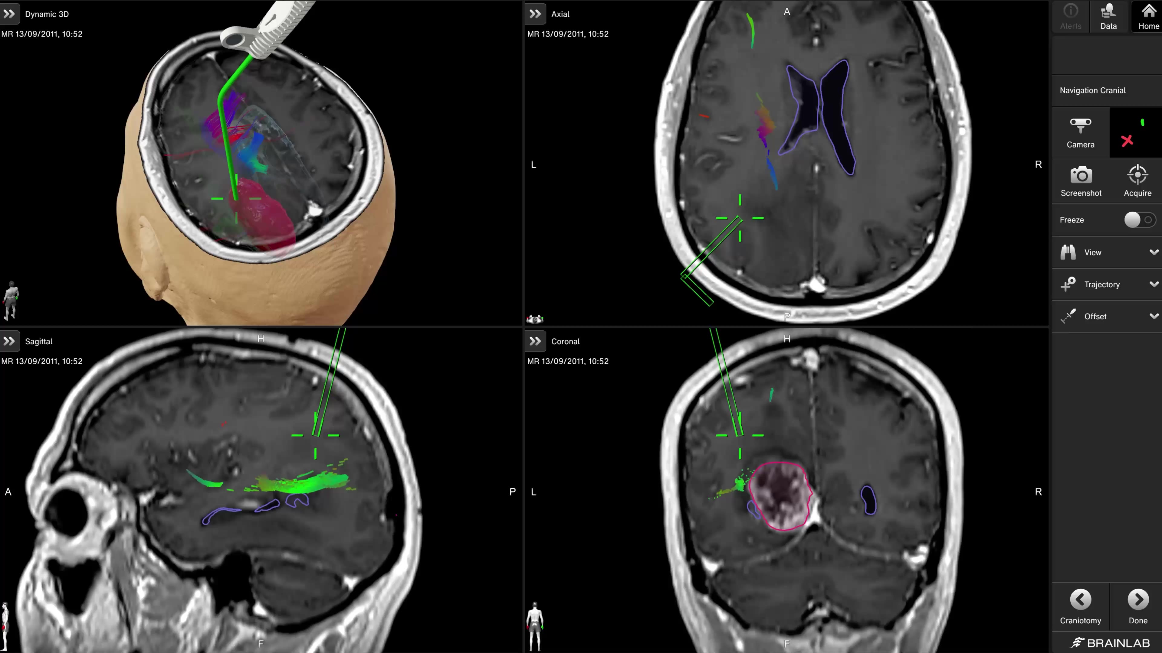
EM NavigationNavigate without constraints
When a case calls for touchscreen-free, pinless navigation—especially for pediatric patients—electromagnetic tracking adds flexibility to the workflow. Cranial EM Navigation supports workflow-guided setup, registration and navigation across a range of patient positions, including challenging ones. Designed for ease of use, an integrated remote control lets surgeons freeze the screen, capture screenshots or acquire a trajectory without touching the display so they can focus and keep the workflow moving.

Microscope NavigationBring clarity into view
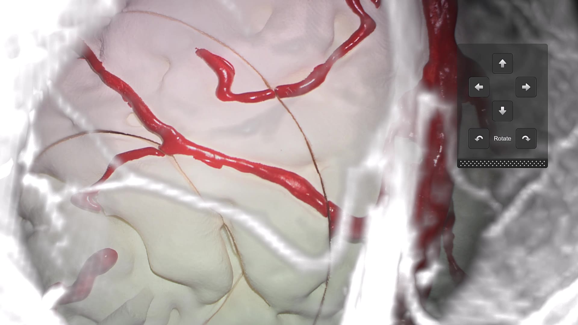
In tumor resection, the microscope is where the most critical decisions happen—and Microscope Navigation ensures that the data you need is always in your line of sight. AR overlays bring tumor boundaries, fiber tracts and critical anatomical structures directly into your surgical view, helping you navigate the margin between tumor and healthy tissue with confidence. Robotic microscope positioning adjusts automatically so you can stay focused on the resection, not the equipment.


Stay true as anatomy changes
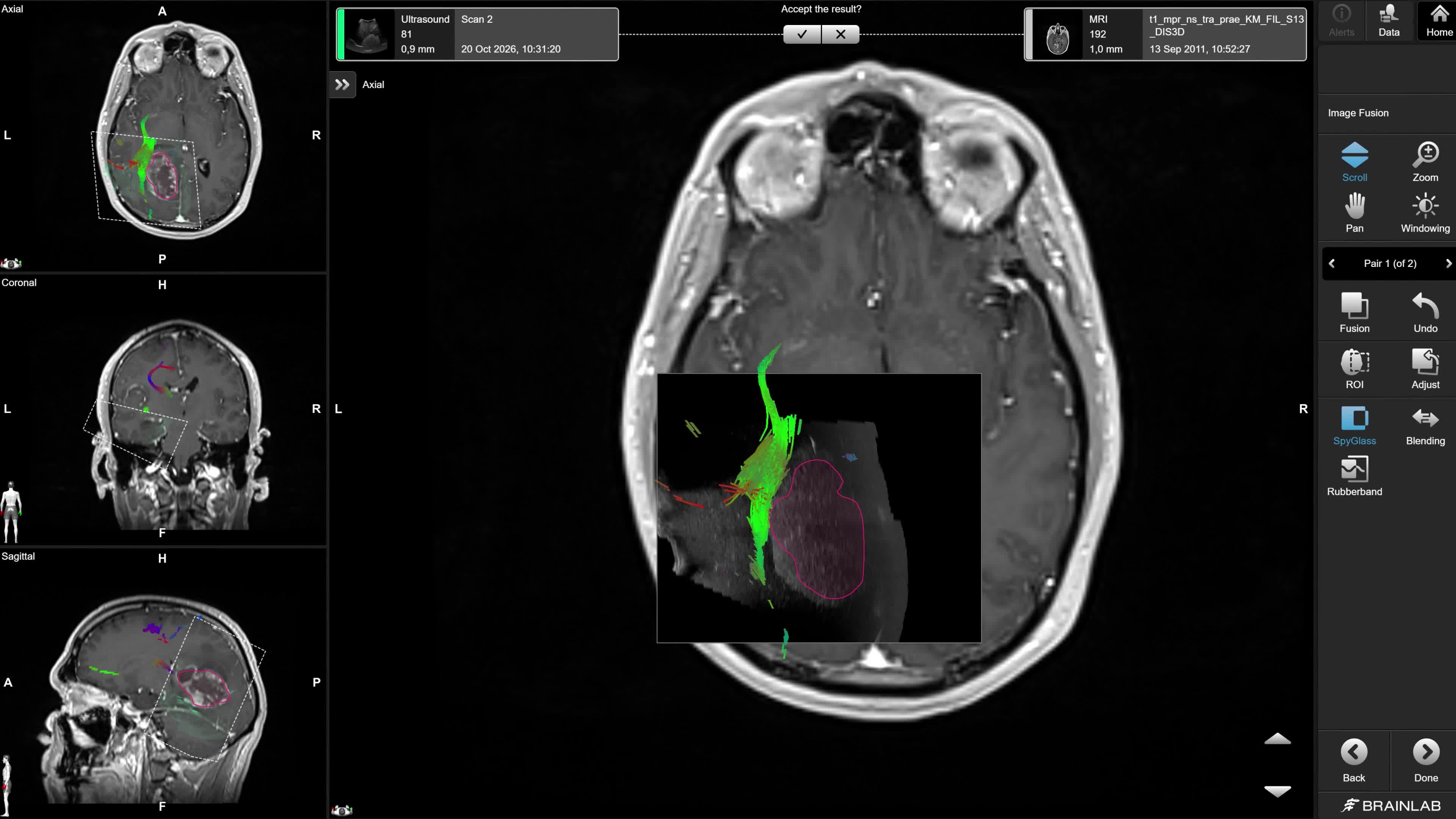
Even the most precise preoperative plan must account for the challenge of brain shift. As the procedure progresses, changes in anatomy or registration can create discrepancies between preoperative images and the surgical field—sometimes without being immediately apparent. The information guiding you should reflect the anatomy in front of you.


Experience innovation up close
See our cranial workflow in action. Request a personalized demo to witness how our solutions can support you in precise tumor resection.
The statements made by the surgeon during this interview represent their personal opinion and experiences. These statements may not be supported by scientific evidence or peer-review research. For verified information about the device, please refer to the manufacturer’s official documentation or consult clinical guidance.
Krieg S. et al., Preoperative motor mapping by navigated transcranial magnetic brain stimulation improves outcome for motor eloquent lesions. Neuro Oncol. first published online February 9, 2014
Not yet commercially available in several countries. Please contact your sales representative.