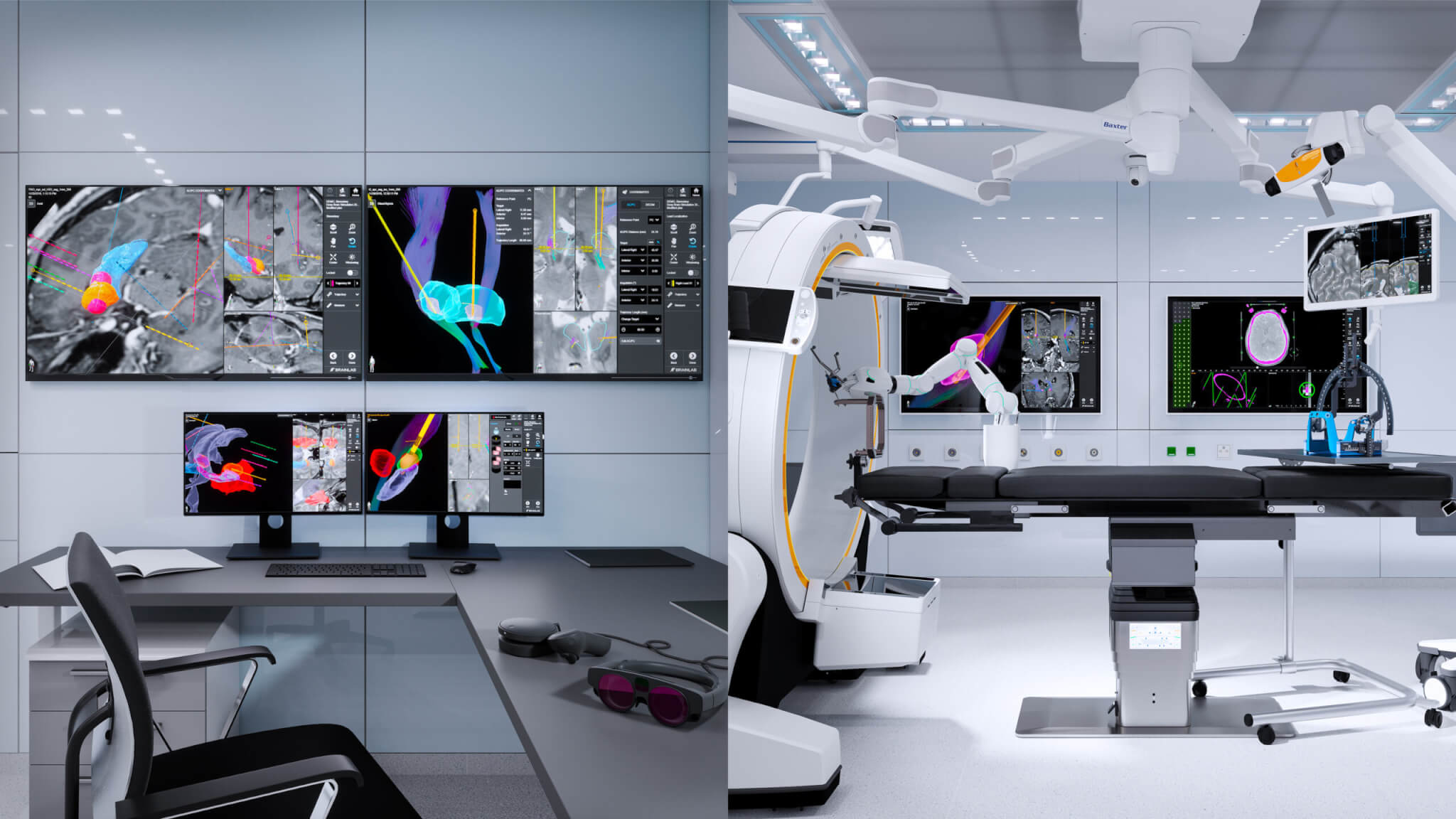
As we look toward the future, our mission is to address the unmet needs of patients suffering from neurological disorders such as Parkinson’s disease, dystonia, essential tremor and drug-resistant epilepsy.
Behind the scenes of functional neurosurgery
Functional neurosurgery is a branch of neurosurgery in which symptoms related to central nervous system disorders, including Parkinson’s disease, dystonia, essential tremor and drug-resistant epilepsy, are addressed by altering the function of specific targets in the brain and the neural circuits within the targets 1.
Origins
In the past, the ability to identify brain targets on patient imaging was highly limited due to a lack of contrast and clear image resolution.
As such, procedures like deep brain stimulation (DBS) historically relied on additional workflow steps such as intraoperative electrophysiology and clinical tests that needed to be conducted while the patient was awake.
Present
In the last few years, advances in imaging technology and novel treatment modalities have considerably changed the field of functional neurosurgery.
Today, imaging guides clinicians in the planning and execution of stereotactic procedures. In parallel, intraoperative imaging technologies are being developed with the goal of making such procedures more efficient and effective.
Future
From accurate brain imaging technology to improved intraoperative navigation solutions, refined therapeutic paradigms and more, the field of functional neurosurgery will continue to rapidly progress.
In the future, functional neurosurgery procedures will further expand into additional neurological and psychiatric indications and become even more accessible to patients.
Our vision at Brainlab is to offer a platform that analyzes, understands and optimizes patient brain functions. By leveraging the Brainlab data ecosystem and the company’s ever-evolving data enrichment capabilities, we seamlessly incorporate different technologies into scalable, streamlined and optimized functional neurosurgery workflows.
Our vision at Brainlab is to offer a platform that analyzes, understands and optimizes patient brain functions. By leveraging the Brainlab data ecosystem and the company’s ever-evolving data enrichment capabilities, we seamlessly incorporate different technologies into scalable, streamlined and optimized functional neurosurgery workflows.
Bring visual clarity and control
into surgery
Our functional focus at Brainlab
Although functional neurosurgery is applicable to a handful of chronic neurological conditions, our focus at Brainlab is using our software and hardware solutions for two overarching topics – deep brain stimulation and the diagnosis and treatment of drug-resistant epilepsy.

Brainlab workflow
and solutions for DBS
Brainlab functional neurosurgery solutions are designed to provide an unparalleled 3D visualization of patient anatomy in relation to deep brain stimulation (DBS) lead contacts from preoperative planning to the start of DBS implantation up through postoperative implant programming.
Learn more

Brainlab workflow
and solutions for epilepsy
Brainlab epilepsy solutions bridge the gaps between hypothesis formulation, advanced planning information, surgical team communication and the navigation of surgical platforms.
Learn more
Access your roadmap to the brain with easy-to-integrate functional neurosurgery software and hardware solutions.
1
https://www.sciencedirect.com/science/article/abs/pii/B9780123970251001184
2
https://www.who.int/news-room/fact-sheets/detail/epilepsy
